-
CVD Graphene Oxygen Sensor for Lung Membranes — UIUC, 2020
May 20, 2026 | ACS MATERIAL LLCKim, M., Porras-Gomez, M., & Leal, C. (2020). Graphene-based sensing of oxygen transport through pulmonary membranes. *Nature communications*.
Nature communications · 2020
UIUC researchers built a CVD-graphene oxygen sensor (ACS Material) revealing how cardiolipin stalks raise O2 permeability in pulmonary membranes.
About this research
Researchers at the University of Illinois at Urbana-Champaign used CVD graphene on copper foil from ACS Material, LLC to fabricate a micrometer-scale oxygen permeation sensor that directly measures gas transport through model pulmonary membranes. Published in Nature Communications (2020), the study by Kim, Porras-Gomez, and Leal combines graphene-based sensing with grazing-incidence small-angle X-ray scattering (GISAXS), confocal fluorescence microscopy, and atomic force microscopy to show that cardiolipin — a mitochondrial lipid overexpressed in bacterial pneumonia — triggers a structural transformation of pulmonary surfactant films that markedly increases oxygen permeability. The result links a molecular-scale lipid rearrangement to a physiologically significant change in alveolar gas exchange.
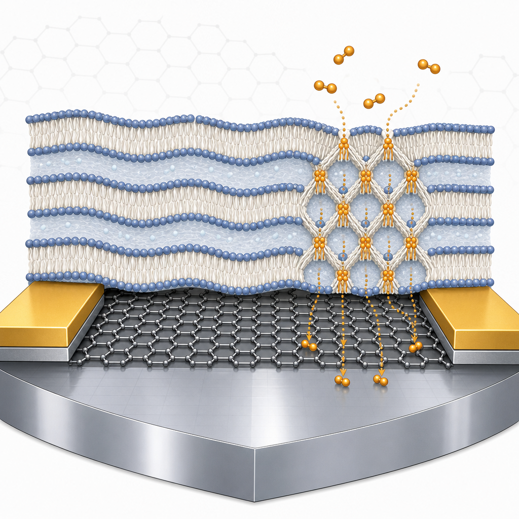
Pulmonary surfactant is a lipid–protein film that coats the alveolar surface and mediates the diffusion of oxygen from inhaled air into the bloodstream. In bacterial pneumonia, the surfactant composition is altered: cardiolipin (CL) accumulates to 5–10 mol%, hydrophilic protein SP-A is depleted, and patients are frequently subjected to hyperoxia therapy. How these compositional changes translate into impaired gas exchange has been difficult to probe because conventional permeability assays lack the spatial resolution and chemical sensitivity required to track oxygen flux through micrometer-thick lipid mesophases under physiologically relevant humidity. The authors address this gap by reading oxygen flux directly as a change in graphene channel resistance, exploiting graphene's exquisite sensitivity to surface-adsorbed gas species and its compatibility with thin-film lipid deposition.
The sensor was fabricated by transferring CVD graphene from ACS Material's graphene/copper foil onto a SiO2/Si wafer. Poly(methyl methacrylate) (PMMA, 950 A2 from MicroChem) was spin-coated as a support layer, the copper was etched, and the PMMA/graphene film was rinsed in DI water before lamination onto the wafer; PMMA was then removed with acetone. A Cr (3 nm) / Au (70 nm) electrode stack was thermally evaporated and photolithographically patterned, after which the graphene was defined into channels by reactive-ion etching (60 sccm O2, 50 W, 60 s). Stock lipid solutions — DPPC, DOPG, cardiolipin, and the clinical surfactant BLES — were spin-coated at 4000 rpm for 30 s directly onto the active graphene area, incubated at 45 °C for 48 h, and stored at 98–100% relative humidity. The graphene channel thus served simultaneously as the substrate on which the lipid mesophase organized and as the transducer reading out oxygen permeation through it.
Oxygen permeability was extracted from real-time two-probe resistance measurements (0.1 V drain bias) in a cryogenic probe station with controlled O2/N2 dosing at >98% RH. Confocal microscopy of Texas Red-DHPE–tagged films verified uniform lipid coverage with virtually no micrometer-scale defects. GISAXS measurements performed in-house and at APS beamline 12-ID-B (14 keV) revealed that healthy DPPC:DOPG (3:1) films and BLES exhibit a smectic lamellar stack with d-spacing near 9.52 nm and a water layer of ~4.5 nm. Adding 3 and 8 mol% cardiolipin (guided by the 5–10 mol% range reported in pneumonia-affected human lungs) shifted the d-spacing to 9.58 nm (dwater ≈ 5.1 nm) and, more importantly, induced periodic protein-free inter-membrane contacts — "stalks" — arranged with rhombohedral symmetry. Thermogravimetry showed water contents of 14.9 wt% and 15.2 wt% for healthy and diseased films, respectively. The graphene sensor recorded a substantially shorter saturation time for oxygen in cardiolipin-containing films, quantifying the structural change as a significant enhancement of oxygen gas permeability. Calcium chloride at ~1 mM (4 mol%) was included to match physiological Ca²⁺ levels (1–5 mM).For biomedical applications like in-vitro cell monitoring, the pristine lattice structure of the sensor is critical. Our CVD Graphene undergoes a highly optimized transfer process to ensure minimal PMMA residue and exceptional biocompatibility. Explore our CVD Graphene Substrates to build your next-generation biosensors.
 These findings reframe a clinical observation in pneumonia — disturbed alveolar gas exchange — as a structurally driven phenomenon: stalk phases in the surfactant film create lower-resistance pathways for oxygen, which combined with hyperoxia therapy may contribute to oxidative injury. The graphene-based platform itself is broadly extensible. Because it tracks gas flux through a deposited soft-matter film with micrometer lateral resolution and high temporal resolution, it can be applied to other respiratory disorders (ARDS, cystic fibrosis surfactant dysfunction), to screening of synthetic surfactant replacements, to drug-delivery studies through lipid barriers, and to fundamental membrane biophysics where dissolved-gas transport is otherwise difficult to quantify. Coupling with synchrotron GISAXS and AFM, as demonstrated here, gives a structure–transport correlation rarely achieved in living-tissue mimics.
For researchers planning similar studies, the central enabling component is high-quality monolayer CVD graphene transferable onto arbitrary device substrates. ACS Material's CVD Graphene on Copper Foil — the exact product used in this work — is available in research and pilot quantities and supports the PMMA-assisted transfer and lithographic patterning workflow described above. Groups working on gas sensing, biosensing, 2D-material biointerfaces, or lipid-membrane biophysics can readily adapt the methodology, and the broader CVD graphene catalog includes formats on SiO2, quartz, and PET for related device architectures.How ACS Material products were used
- CVD Graphene on Copper Foil (CVD Graphene) — “graphene/copper foil was purchased from ACS Material, LLC (Pasadena, CA)”
Product Performance in this Study
The CVD graphene on copper foil was transferred onto SiO2/Si wafers and patterned to form the active sensing channel of a micrometer-scale oxygen permeability sensor. The graphene channel provided the resistive response used to quantify oxygen transport through pulmonary lipid membranes deposited directly on top of it, enabling reproducible measurements at high humidity.
Related product categories
Frequently asked questions
How is CVD graphene used to measure gas permeability through lipid membranes?
A monolayer of CVD graphene is transferred onto a SiO2/Si wafer, patterned into a channel with metal electrodes, and coated with the lipid film under study. Because graphene's resistance is highly sensitive to adsorbed gas molecules, the time required for the channel resistance to saturate under O2 dosing serves as a quantitative measure of how quickly oxygen permeates the overlying lipid layer at controlled humidity.
Why does cardiolipin increase oxygen permeability in pulmonary surfactant?
GISAXS data show that adding 3–8 mol% cardiolipin to DPPC:DOPG or BLES films promotes periodic protein-free inter-membrane contacts called stalks, arranged with rhombohedral symmetry. These stalks bridge adjacent bilayers and create lower-resistance pathways for dissolved oxygen, so films containing cardiolipin saturate the graphene sensor with O2 much faster than healthy lamellar films of the same composition.
What CVD graphene format is suitable for fabricating thin-film gas sensors?
Monolayer CVD graphene grown on copper foil is the standard starting format. A PMMA support layer is spin-coated, the copper is etched, and the PMMA/graphene stack is transferred to SiO2/Si or another target substrate before PMMA removal. The transferred graphene can then be defined into channels by photolithography and reactive-ion etching, as demonstrated in this Nature Communications study using ACS Material graphene/copper foil.