
Fresh meat and spoiled meat can look almost identical — but at the molecular level they are not. As bacteria break down the energy molecules in muscle tissue, they leave behind a chemical fingerprint of decay, and the clearest marker of all is xanthine. A team at the University of Florida built a small electrochemical sensor that reads that marker directly, coating its electrode with graphene oxide from ACS Material to turn rising xanthine into a rising electrical current. The result is a fast, low-cost route to a number the meat industry has always wanted at the point of use: how fresh, exactly, is this sample? This article walks through how the device works, how the graphene-oxide layer is built, what it measured, and where the same platform goes next.
Vanegas, D. C.; Gomes, C.; McLamore, E. S. Xanthine Oxidase Biosensor for Monitoring Meat Spoilage. Proc. SPIE 9107, Smart Biomedical and Physiological Sensor Technology XI, 2014. DOI: 10.1117/12.2050489
University of Florida researchers (with collaborators at Texas A&M University and Universidad del Valle) built a xanthine oxidase biosensor on a platinum–iridium electrode using graphene oxide supplied by ACS Material, reaching a 150 nM detection limit for the spoilage marker xanthine.
Short answer: meat spoilage releases xanthine, and the amount of xanthine tracks how far decay has progressed. The University of Florida sensor measures it by stacking catalytic layers on a platinum–iridium electrode tip — platinum-black nanoclusters, a reduced-graphene-oxide/nanoceria film made in situ from ACS Material graphene oxide, a second platinum-black layer, and finally the enzyme xanthine oxidase held in a hydrogel. The reduced graphene oxide is the conductive scaffold that ties the whole stack together. The finished sensor detects xanthine down to 150 nM, responds in about 5 seconds, and still works after a week of continuous use — the three things a point-of-use freshness meter actually needs.
Why meat freshness comes down to xanthine
When an animal’s muscle stops getting oxygen, the energy molecule ATP that powered it begins to break down. Enzymes and bacteria carry that breakdown forward along a fixed chemical chain — ATP to ADP to AMP, then to inosine monophosphate, inosine, hypoxanthine, and finally xanthine. The further spoilage has gone, the more hypoxanthine and xanthine have accumulated. The meat industry captures this in a single number called the K-value, the ratio of these late-stage breakdown products to all the ATP-related compounds present; a low K-value means fresh, a high K-value means spoiled1.
The problem is measuring it. The standard K-value assay relies on chromatography — accurate, but slow, expensive, and tied to a laboratory. None of that fits a slaughterhouse line, a distribution dock, or a retail cold case, where a decision about a batch of product has to be made in seconds. A sensor that could read hypoxanthine or xanthine directly, cheaply, and on the spot would let processors and inspectors flag contaminated product before it reaches a consumer. That is the gap this device set out to fill: an electrochemical biosensor specific to xanthine, fast enough and rugged enough for the field.
See it: as meat ages, the current climbs
The sensing idea is easier to see than to describe. In the interactive below, drag the slider to advance the storage time. As the simulated meat ages it releases more xanthine; the graphene-oxide electrode oxidizes that xanthine, and the current trace on the right rises from a low, flat baseline toward a high “spoiled” signal. The freshness label and current readout update with it. It is a schematic of the mechanism, not real sensor data, but it captures the central relationship the device exploits: more spoilage → more xanthine → more current.
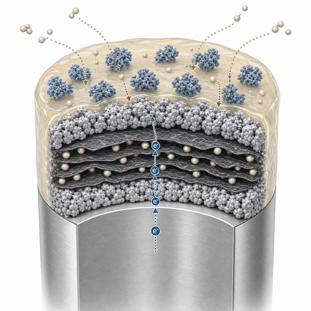
How the graphene-oxide “sandwich” electrode is built
The heart of the sensor is a layered “sandwich” of nanomaterials built directly onto the tip of a platinum–iridium working electrode just 1.6 mm across. Each layer has a job, and ACS Material graphene oxide is the ingredient that makes the central layer work.
The team first decorated the bare Pt/Ir tip with amorphous platinum nanoclusters by sonoelectrodeposition — depositing platinum from a chloroplatinic-acid solution while applying ultrasound — to create a rough, catalytically active base. Separately, they prepared the key transduction layer by mixing a cerium-oxide (nanoceria) dispersion, the ACS Material graphene oxide, and L-ascorbic acid in a 1 mL : 2 mg : 8 mg ratio and ultrasonicating for 30 minutes. During that step the ascorbic acid chemically reduces the graphene oxide into reduced graphene oxide (rGO) right around the nanoceria particles — an in situ reduction that builds the conductive composite in one pot. A 2 µL drop of this CeO/rGO suspension was spin-coated onto the platinized tip at 2600 rpm, a second platinum-black layer was deposited on top, and the stack was capped with the enzyme xanthine oxidase encapsulated in laponite clay hydrogel and a silica sol-gel.
Why these particular materials together? The three inorganic components are complementary, an approach the same group has explored in related nanoceria–platinum–graphene biosensors2. The platinum-black nanoclusters supply catalytic junctions and surface roughness; the nanoceria boosts catalysis of the hydrogen peroxide and superoxide that oxidase enzymes generate; and the reduced graphene oxide is the continuous conductive scaffold that bridges the platinum junctions and physically supports the catalytic ceria. Holding the enzyme in a hydrogel matrix, rather than binding it directly to the metal, is a well-established way to keep it active and stable on the electrode3. The net architecture is an nPt–rGO/CeO–nPt sandwich with the biorecognition enzyme on top.
What the sensor achieved
Cyclic voltammetry in a ferricyanide solution confirmed that the modified electrode supported clean, reversible redox chemistry. Applying the Randles–Sevcik relationship, the team calculated the electroactive surface area of the finished probe at 0.062 cm² — about a 226% increase over the bare Pt/Ir tip. Electron microscopy backed this up, showing the platinum/rGO/ceria stack formed a rough, high-area topography in intimate contact with the solution. More surface area means more sites where the reaction can happen, which is exactly what a sensitive amperometric sensor needs.
Run as a detector — held at +500 mV versus a silver/silver-chloride reference — the electrode produced a clean, stepwise jump in current with each addition of xanthine, yielding a straight calibration line. The headline performance figures are summarized below1.
| Performance metric | Reported value |
|---|---|
| Sensitivity | 2.14 ± 1.48 µA per mM |
| Response time | 5.2 ± 1.5 seconds |
| Lower detection limit | 150 ± 39 nM xanthine |
| Electroactive surface area | 0.062 ± 0.008 cm² (about 226% above a bare tip) |
| Operating potential | +500 mV vs. Ag/AgCl |
| Operational stability | retained at least 88% of activity after 7 days of continuous use |
| Main interferent | ascorbic acid (addressed with a Nafion overlayer) |
The 150 nM detection limit is the figure that matters most: it sits well below the xanthine concentrations associated with even early-stage spoilage, so the sensor can flag a problem long before meat is visibly or odorously “off.”
Selectivity and week-long stability
A field sensor has to ignore the things it is not measuring and keep working for more than a single afternoon. On selectivity, the team challenged the electrode with urea, glucose, and ascorbic acid; only ascorbic acid (vitamin C) produced significant interference, and the authors note the standard fix — a thin Nafion ion-exchange layer that blocks negatively charged ascorbate while letting the target through. On durability, the device retained at least 88% of its initial sensitivity after seven days of continuous operation, evidence that the laponite-and-sol-gel matrix successfully stabilized the xanthine oxidase against the gradual loss of activity that plagues many enzyme electrodes. Fast response, sub-micromolar sensitivity, and week-long stability are the three attributes an in-line freshness screen most needs, and this device delivered all three at once.
A platform beyond meat
The most useful thing about this work is that the chemistry is not specific to meat. Xanthine oxidase belongs to a large family of oxidase enzymes, and every one of them generates hydrogen peroxide when it acts on its target molecule. The nanoceria/rGO/platinum-black stack is, in effect, a general-purpose transducer for sensing that hydrogen peroxide — so swapping the enzyme on top turns the same electrode into a sensor for something else entirely. The same group has extended closely related electrodes to real-time monitoring of metabolite transport in living systems4, and the broader opportunity spans glucose monitoring, lactate sensing for sports physiology, neurotransmitter detection, and on-chip bioreactor control. The authors’ own next steps were practical ones: integrate the Nafion layer to suppress the ascorbic-acid interference, and miniaturize the design toward disposable, single-use probes.
For a research group, the deeper lesson is about the starting material. Rather than synthesizing graphene oxide in-house — a multi-step, hazardous process — the team bought a consistent commercial graphene oxide and reduced it to rGO directly on the electrode. That turns a difficult synthesis into a simple mixing-and-coating step, and it makes the result reproducible from batch to batch.
The ACS Material graphene oxide used
The study used graphene oxide from ACS Material as the matrix for its central transduction layer, reducing it in situ with ascorbic acid into the conductive rGO scaffold that ties the sandwich together. Graphene oxide is the workhorse of this kind of electrode chemistry precisely because its surface carries abundant, uniform oxygen groups — hydroxyl, epoxy, and carboxyl — which make it disperse readily in water, reduce cleanly to rGO, and anchor nanoparticles and enzymes. If you are new to the material, our complete guide to graphene oxide and the wider graphene family covers how it is made and why those oxygen groups matter.
ACS Material offers graphene oxide in several formats suited to nanocomposite electrode work in food-safety, biomedical, and environmental sensing — including single-layer graphene oxide powders and ready-to-use graphene oxide dispersions, alongside the full Graphene Series.
The success of GO-based enzymatic biosensors heavily depends on the abundant and uniform oxygen functional groups on the graphene surface. Whether you are developing food safety diagnostics or wearable health monitors, consistent material quality is your baseline. Explore our highly stable Graphene Oxide Dispersions and Powders to scale your biosensor prototypes with confidence.
Frequently asked questions
How is graphene oxide used in a xanthine biosensor for meat spoilage?
Graphene oxide is mixed with cerium-oxide nanoparticles and ascorbic acid and ultrasonicated, so the ascorbic acid reduces the graphene oxide to reduced graphene oxide (rGO) in situ around the nanoceria. That suspension is spin-coated onto a platinum–iridium electrode already coated with platinum black, a second platinum-black layer is added, and the enzyme xanthine oxidase — held in a laponite hydrogel — is placed on top. The rGO acts as the conductive scaffold that links the catalytic layers.
What detection limit can this graphene-oxide xanthine biosensor reach?
The device reached a lower detection limit of 150 ± 39 nM xanthine, with a sensitivity of 2.14 ± 1.48 µA/mM and a response time of about 5 seconds. It retained at least 88% of its activity after seven days of continuous use — well within the range needed to flag early-stage bacterial spoilage in food-safety screening.
Why combine reduced graphene oxide with nanoceria and platinum black?
The three materials are complementary. Platinum-black nanoclusters create catalytic junctions and surface roughness; reduced graphene oxide bridges those junctions as a continuous conductive scaffold; and nanoceria boosts catalysis of the hydrogen peroxide that oxidase enzymes produce. Together they raised the electroactive surface area by about 226% over a bare electrode and enabled fast, low-noise amperometric detection.
Can the same sensor design detect things other than meat spoilage?
Yes. Any oxidase enzyme generates hydrogen peroxide, so the nanoceria/rGO/platinum-black stack works as a general transducer — swapping the surface enzyme adapts it to targets such as glucose, lactate, or neurotransmitters, as well as bioprocess and environmental monitoring.
References
This article is provided by ACS Material LLC for educational purposes and describes a published study that used graphene oxide in a xanthine oxidase biosensor for meat-spoilage monitoring. The performance values cited — a 150 nM detection limit, 2.14 µA/mM sensitivity, a roughly 5-second response, a ~226% increase in electroactive area, and 88% activity retention over seven days — refer to the specific electrodes, materials, and conditions reported in the referenced work; results obtained with other grades, formats, or fabrication conditions will differ. Consult product datasheets and safety data sheets for grade-specific specifications and handling guidance. The interactive simulator is a schematic teaching tool that illustrates the qualitative relationship between meat spoilage, xanthine release, and sensor current; it is not real sensor data or predictive software, and actual sensor performance must be established experimentally.