-
Black Phosphorus Nanosheets for Bone Scaffolds — Mayo Clinic, 2019
Jun 11, 2026 | ACS MATERIAL LLCLiu, X. et al. (2019). Two-dimensional black phosphorus and graphene oxide nanosheets synergistically enhance cell proliferation and osteogenesis on 3D printed scaffolds. *ACS Applied Materials & Interfaces*. https://doi.org/10.1021/acsami.9b04121
ACS Applied Materials & Interfaces · 2019
Mayo Clinic researchers used ACS Material black phosphorus powder to functionalize 3D-printed PPF scaffolds, synergistically boosting osteogenesis with graphene oxide.
About this research
Researchers at the Mayo Clinic, led by Lichun Lu, used black phosphorus (BP) powder purchased from ACS Material, LLC (Pasadena, CA) to fabricate 2D BP nanosheets that, when combined with graphene oxide (GO), synergistically enhanced pre-osteoblast proliferation, collagen production, biomineralization, and osteogenic differentiation on 3D-printed poly(propylene fumarate) (PPF) scaffolds. Published in ACS Applied Materials & Interfaces (2019), the study compares scaffolds coated with BP alone, GO alone, and a combined GO@BP layer, showing that the dual coating outperforms either single 2D material across nearly every metric relevant to bone tissue engineering.
Bone regeneration on synthetic scaffolds depends on a delicate balance of surface topography, protein adsorption, cell adhesion, and the local release of osteogenic ions. Polymer scaffolds such as PPF are biocompatible and 3D-printable but lack intrinsic bioactivity. Strategies to functionalize their surfaces with 2D nanomaterials have attracted growing attention because graphene derivatives can boost cell adhesion through surface chemistry, while black phosphorus uniquely releases phosphate ions that are biocompatible and directly relevant to hydroxyapatite formation. Combining these two materials addresses an open challenge in orthopedic scaffold design: how to integrate stable adhesion-promoting layers with continuously bioactive ion-releasing nanostructures in a single, uniform coating compatible with stereolithography-printed polymer constructs.
In the study, 30 mg of BP powder (ACS Material, LLC) was dispersed in 30 mL of deionized water and sonicated for 1 h with a probe sonicator, then for 12 h with an ice-bath sonicator to liquid-exfoliate few-layer BP nanosheets. After centrifugation, the supernatant was lyophilized to yield BP nanosheet powder. AFM confirmed sheet thicknesses of 16–28 nm and lateral sizes peaking near 4 µm. GO was synthesized by an improved Hummers' method. PPF scaffolds were 3D-printed by stereolithography (5 × 5 × 5 mm cubes with 1 mm pores and 0.5 mm ridges), aminated by hexamethylenediamine ammonolysis, then coated by soaking in BP, GO, or GO@BP solutions (1 mg/mL each). SEM and EDS mapping confirmed that BP nanosheets were homogeneously wrapped by GO, with phosphorus signals stronger on GO@BP scaffolds than on BP-only scaffolds—indicating that GO improved BP retention on the scaffold surface.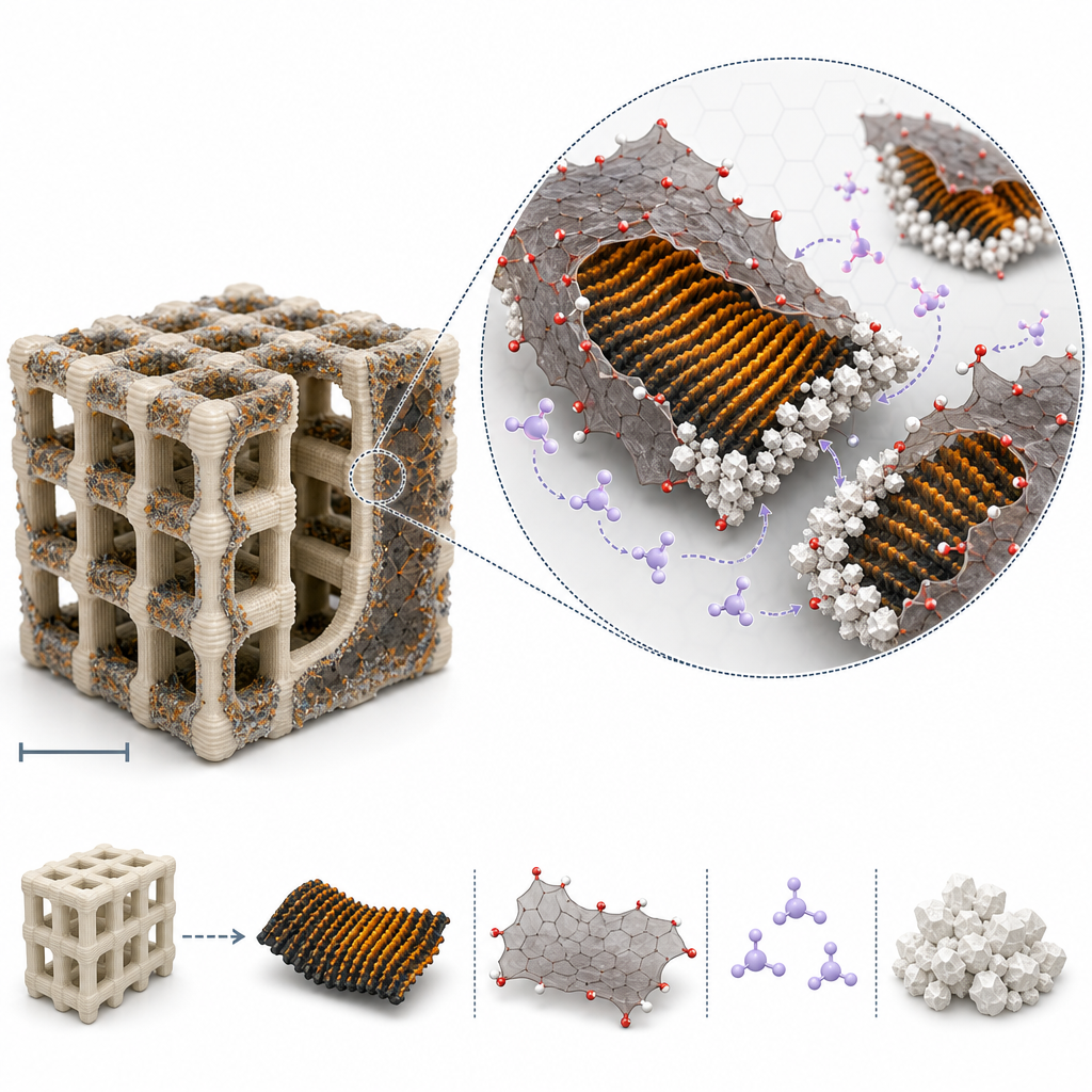
Key results highlight the synergy of the two 2D materials. AFM root-mean-square roughness was significantly higher on BP-coated than on GO-coated scaffolds, and highest on GO@BP scaffolds. Protein adsorption from FBS-supplemented α-MEM was significantly elevated for GO and GO@BP groups versus PPF-Amine and BP groups. EDS quantified more phosphorus on GO@BP than on BP-only scaffolds, and phosphate-release assays at 37 °C showed sustained release from BP scaffolds plateauing by day 10, while GO@BP scaffolds released higher cumulative phosphate, with the GO layer slowing the rate after day 10 by limiting oxidation of buried BP. MC3T3 pre-osteoblasts cultured on GO@BP scaffolds reached the highest cell densities at days 1, 3, and 6, with cell-attachment rates also highest for GO-containing scaffolds. Total collagen production after 1 week was greatest on GO@BP scaffolds. After 7 days in simulated body fluid, GO@BP scaffolds developed the thickest mineral coverage, with calcium-phosphate aggregates nucleating along BP nanosheet edges. Osteogenic markers showed BP significantly elevated ALP activity in both standard and β-GP/AA media, while OCN content at 21 days was highest on GO@BP scaffolds in both media. Leaching-medium cytotoxicity assays through 7 days showed no cytotoxic effect.
These results have direct implications for orthopedic implants, craniofacial reconstruction scaffolds, and broader bone tissue engineering platforms where surface bioactivity must be added to inert printable polymers. The GO@BP coating concept could be adapted to other photo-cured or extruded scaffolds—including PLA, PCL, and PEG-based hydrogels—and may translate to nerve guidance conduits and cardiac patches where 2D-material-mediated charge transport and ECM mimicry are useful. The paper itself points toward future in vivo evaluation of GO@BP-functionalized scaffolds and to combinations of 2D materials with growth-factor delivery for accelerated bone repair, suggesting a modular platform for ion-releasing biomaterials.
For researchers pursuing similar work, the black phosphorus powder used here is part of ACS Material's graphene-like 2D materials catalog, alongside MoS2, WS2, and h-BN products. The study demonstrates that simple liquid exfoliation of commercially available BP powder is sufficient to produce nanosheets active in mineralization and osteogenesis, lowering the barrier for biomedical groups without specialized 2D-material synthesis capabilities. The reproducibility of the exfoliation protocol and the consistency of the reported phosphate-release and ALP data underscore the practical value of starting from well-characterized BP starting material for tissue-engineering studies.How ACS Material products were used
- Black Phosphorus Powder (Graphene-like Materials) — “30 mg of BP powder (ACS Material, LLC, Pasadena, CA) was mixed with 30 mL deionized (DI) H2O and sonicated for 1 hour using a probe model sonicator”
Product Performance in this StudyThe ACS Material black phosphorus powder served as the precursor for liquid-exfoliated 2D BP nanosheets used to functionalize 3D-printed scaffolds. The exfoliated nanosheets contributed phosphate ion release, enhanced surface roughness, accelerated biomineralization, and significantly elevated ALP activity in pre-osteoblasts, demonstrating high performance for the intended osteogenic application.
Related product categories
Frequently asked questionsHow do black phosphorus nanosheets enhance osteogenesis on 3D-printed scaffolds?
Black phosphorus nanosheets continuously release phosphate ions in aqueous environments, which raises local phosphate concentration and promotes calcium-phosphate biomineralization on scaffold surfaces. The released phosphate also up-regulates alkaline phosphatase activity in pre-osteoblasts, accelerating osteogenic differentiation. Additionally, BP nanosheets increase surface roughness, providing topographical cues that improve focal adhesion development and cell elongation, both essential for early bone-tissue regeneration.
Why combine graphene oxide with black phosphorus for tissue engineering scaffolds?
Graphene oxide and black phosphorus play complementary roles. GO carries negative surface charges and large surface area, which strongly enhance protein adsorption and pre-osteoblast attachment. BP releases phosphate ions that drive mineralization and ALP activity. Wrapping BP nanosheets with GO also improves their retention on the scaffold surface and reduces oxidative degradation, yielding higher cumulative phosphate release and the strongest osteogenic response observed in the study.
How are 2D black phosphorus nanosheets prepared from commercial BP powder?
Commercial bulk black phosphorus powder is typically liquid-exfoliated. The Mayo Clinic team dispersed 30 mg of BP powder in 30 mL deionized water, applied probe sonication for 1 hour, followed by 12 hours of ice-bath sonication. Centrifugation removed unexfoliated material and the supernatant was lyophilized to recover nanosheet powder. AFM confirmed thicknesses of 16–28 nm and lateral sizes around 4 µm, suitable for surface coating applications.