-
Carboxyl Graphene Oxide TERS Imaging - Hangzhou Dianzi University, 2018
May 26, 2026 | ACS MATERIAL LLCSu, W. et al. (2018). In situ topographical chemical and electrical imaging of carboxyl graphene oxide at the nanoscale. *Nature communications*.
Nature communications · 2018
Researchers used carboxyl graphene oxide from ACS Material to demonstrate ~10 nm TERS mapping of defects and functional groups, correlating Fermi level with defect density.
About this research
Researchers at Hangzhou Dianzi University, in collaboration with the National Physical Laboratory (UK), Utrecht University, and HORIBA Scientific, used carboxyl-functionalized graphene oxide (GO–COOH) supplied by ACS Material to demonstrate the first combined topographical, chemical and electrical nanoscopy of a functionalized two-dimensional carbon surface, achieving approximately 10 nm spatial resolution. Published in Nature Communications in 2018, the study integrated tip-enhanced Raman spectroscopy (TERS) with Kelvin probe force microscopy (KPFM) on the same sample area using the same Au-coated probe, and revealed an inverse correlation between local defect density (ID/IG ratio) and the Fermi level on the GO–COOH surface. The result establishes a non-destructive, multi-parameter framework for characterizing optoelectronic devices built on 2D materials under realistic operating conditions.
Functionalized graphene oxide is one of the most widely investigated 2D materials, with applications spanning lithium-ion battery anodes, hybrid solar cells, fuel cell electrodes, biosensors, and targeted drug delivery. Its performance depends critically on the spatial distribution of carboxyl, carbonyl, hydroxyl, epoxide and methyl groups, as well as the population of lattice defects that arise during oxidation and reduction steps. Conventional techniques struggle to resolve this chemistry at the relevant length scale: X-ray photoelectron spectroscopy operates at millimetre resolution, confocal Raman is limited to 200–300 nm, and aberration-corrected TEM yields topographic detail but limited molecular fingerprinting. A method that simultaneously delivers chemical specificity, topographic information, and electronic-structure data at the nanoscale has therefore been a longstanding goal for the graphene-oxide community and for 2D-materials device engineering more broadly.
The ACS Material carboxyl graphene oxide was used as the central sample under study. As described in the Methods section, “GO–COOH samples for TERS measurements were prepared by spin-coating GO–COOH (ACS Material, USA) on a Au coated glass substrate.” The gold-coated substrate established the gap-mode plasmonic configuration required for tip enhancement. The deposited flakes formed both few-layer (1–2 layers, ~1–2 nm thick) and thicker (≈5-layer) regions visible in AFM topography. TERS measurements employed an OmegaScope AFM coupled to an XploRA Raman spectrometer (HORIBA Scientific) in side-illumination geometry with a 638 nm excitation laser, a 100× 0.7 NA long working distance objective, and Au-coated AFM TERS tips (k = 7 N/m, f = 150 kHz). Spectra were acquired in SpecTop™ mapping mode at 0.05–0.5 s integration per pixel. Prior microchemical confirmation of the GO–COOH composition was provided by complementary XPS, FTIR and confocal Raman characterization.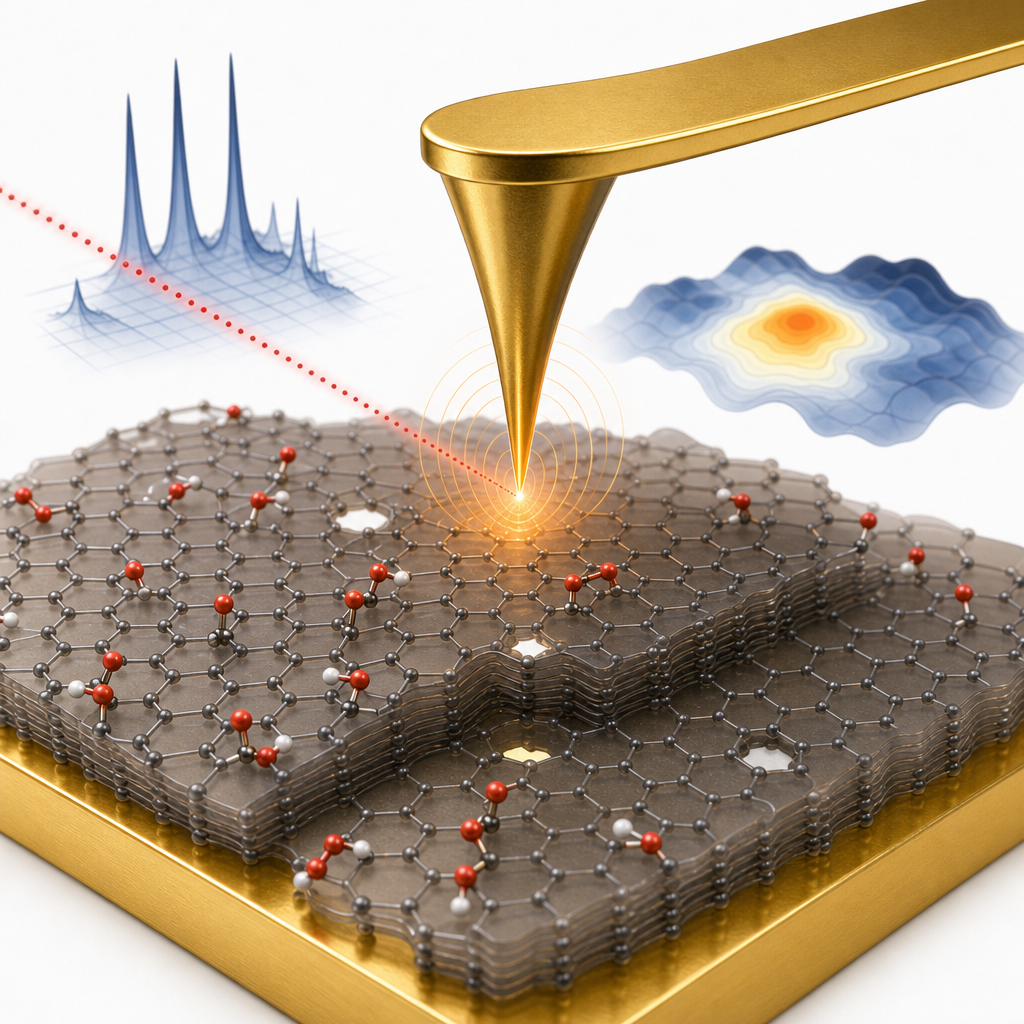
Quantitatively, TERS maps of 100×100 pixel arrays at 10 nm pixel size resolved the D band (1350 cm⁻¹) and G band (1590 cm⁻¹) together with six functional-group bands at 1097, 1179, 1330, 1420, 1654 and 1747 cm⁻¹ assigned to C–O, C–O–C, C–CH3, C–H, C=O and COOH respectively. Gaussian fitting of line profiles across sharp features yielded an average spatial resolution of 10.5 ± 1.7 nm, confirmed across three independent TERS tips. Surface area fractions for C–O, C–O–C, C=O and COOH were measured at 1.3%, 1.0%, 0.5% and 0.6%, while C–CH3 and C–H groups showed broader coverage, with C–CH3 preferentially decorating flake edges and steps. Nanoscale vacancies of 100–600 nm² were identified by the disappearance of D and G bands. KPFM mapping across a central flake measured contact potential differences ranging from −87 to +34 meV, translating to Fermi-level variations of 5.01–5.13 eV (referenced to gold at 5.1 eV). Averaging TERS spectra over eight ~0.012 µm² regions revealed that areas with ID/IG < 1 displayed CPD near −20 meV (Ef ≈ 5.08 eV), while regions with ID/IG > 1.3 dropped to CPD ≈ −80 meV (Ef ≈ 5.02 eV) — an inverse correlation distinct from previously reported α-beam irradiated graphene.
The combined TERS-KPFM workflow has direct implications for optimizing optoelectronic devices that depend on charge injection and transport across functionalized 2D interfaces, including hole-transport layers in polymer solar cells, hybrid photovoltaics, electrochemical biosensors based on RNA-aptamer or peptide conjugates, and graphene-oxide-based drug delivery systems where surface chemistry governs loading and release. The authors specifically highlight applicability to single-layer MoS₂ and other emerging 2D semiconductors, and the methodology lends itself to in-operando measurements on working devices. Future work pointed to in the paper includes deeper analysis of how two-step carboxyl-conversion synthesis routes shape the spatial distribution of residual epoxy and hydroxyl groups.
For researchers pursuing analogous nanoscale characterization, surface-functionalization, or device-fabrication studies, the carboxyl graphene oxide supplied by ACS Material — along with related products in the graphene oxide and graphene series — provides a well-defined starting material with reproducible Raman signatures suitable for advanced microscopy. Its compatibility with spin-coating, gold-substrate deposition and tip-enhanced techniques makes it a practical choice for academic groups developing biosensing platforms, energy-storage electrodes, and optoelectronic test structures. The paper's results are reported objectively; the material performed as required for the demonstration of 10 nm chemical resolution and Fermi-level correlation without further claim of superiority.How ACS Material products were used
- Carboxyl Graphene (-COOH) (Graphene Series) — “GO–COOH samples for TERS measurements were prepared by spin-coating GO–COOH (ACS Material, USA) on a Au coated glass substrate.”
Product Performance in this StudyThe carboxyl-functionalized graphene oxide from ACS Material served as the central sample for nanoscale TERS and KPFM characterization. The material provided sufficient surface functionalization heterogeneity to enable detection of multiple functional groups (C–O, C–O–C, C=O, COOH, C–CH3, C–H) and structural defects at ~10 nm spatial resolution, enabling the study's key correlation between local defect density and Fermi level.
Related product categories
Frequently asked questionsWhat spatial resolution can tip-enhanced Raman spectroscopy achieve on carboxyl graphene oxide?
On carboxyl graphene oxide (GO–COOH) spin-coated onto a gold substrate, the authors achieved an average TERS spatial resolution of 10.5 ± 1.7 nm, verified using Gaussian fits to line profiles across sharp features and reproduced with three independent Au-coated AFM TERS tips. This is roughly 20-30× better than confocal Raman and enables direct mapping of individual defect clusters and functional-group domains within a single GO–COOH flake.
How does local defect density influence the Fermi level of carboxyl graphene oxide?
Combined TERS and Kelvin probe force microscopy measurements on GO–COOH show an inverse correlation between the ID/IG defect ratio and the surface Fermi level. Regions with ID/IG below 1 exhibit CPD near −20 meV (Fermi level ≈ 5.08 eV), while regions with ID/IG above 1.3 fall to CPD ≈ −80 meV (Fermi level ≈ 5.02 eV). This trend differs from α-beam irradiated graphene, reflecting different defect chemistries.
Why is carboxyl-functionalized graphene oxide used in biosensing and drug delivery?
Carboxyl groups on GO–COOH provide reactive handles for covalent coupling to RNA aptamers, peptides, and anticancer drugs, enabling targeted sensing and controlled release. The paper notes applications in theophylline sensing, Caspase-3 protease imaging, and mixed anticancer drug delivery. Mapping the heterogeneous distribution of COOH and other groups at nanoscale, as demonstrated by TERS, helps engineers optimize loading capacity and binding selectivity in such biomedical platforms.