-
Carboxyl Graphene Oxide ATP Aptamer Sensor — Central South University, 2014
Jun 19, 2026 | ACS MATERIAL LLCLiu, Z. et al. (2014). Intracellular detection of ATP using an aptamer beacon covalently linked to graphene oxide resisting nonspecific probe displacement. *Analytical Chemistry*. https://doi.org/10.1021/ac503358m
Central South University · Analytical Chemistry · 2014
Researchers at Central South University covalently linked an ATP aptamer beacon to carboxyl graphene oxide from ACS Material for intracellular ATP imaging.
About this research
Researchers at Central South University, working with collaborators at the University of Waterloo, used carboxyl graphene oxide (Carboxyl GO) supplied by ACS Material to build a covalently linked aptamer beacon that detects ATP inside living cancer cells while resisting the non-specific probe displacement that plagues conventional graphene-oxide aptasensors. By coupling an amino-modified, FAM-labeled ATP aptamer to the carboxyl groups on the GO surface through EDC/NHS chemistry, the team produced a stable fluorescent sensor that turns on selectively in the presence of intracellular ATP. The work, published in Analytical Chemistry in 2014, demonstrates that covalent attachment, rather than physisorption, is the key to reliable biological measurements.
Graphene-oxide-based fluorescent aptasensors are popular because GO efficiently quenches dye-labeled DNA adsorbed on its surface, and target binding releases the DNA to recover the signal. However, in real biological media the same release can be triggered by serum proteins, cellular DNA, or other adsorbents, producing false-positive signals that limit intracellular use. This paper addresses that long-standing challenge in nucleic-acid biosensing by showing that a covalent tether constrains the aptamer to remain on the GO surface even in protein-rich environments, while still permitting the conformational change required for target recognition. The approach is broadly relevant to aptamer-based imaging probes, intracellular small-molecule detection, and any GO-DNA hybrid platform deployed in complex biological samples.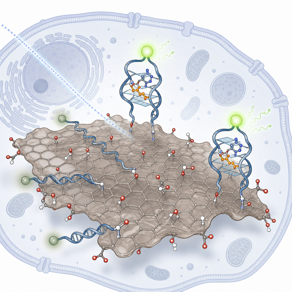
In the methodology, carboxyl GO from ACS Material was dispersed at 10 mg/mL in water by pulsed ultrasonication at 40 W. A 100 μL aliquot was activated with EDC (250 mM) and NHS-sulfate (250 mM) in MES buffer (125 mM, pH 6.0), then reacted with 20 μM amino-modified, FAM-labeled aptamer for 3 h at room temperature in the dark. After centrifugation at 13,500 rpm, physisorbed DNA was stripped off by hybridization with a complementary cDNA, followed by urea (12 M, 70 °C) treatment to remove the cDNA, leaving only covalently bound probes. Coupling efficiency was quantified by measuring supernatant fluorescence in pH 8.5 Tris buffer on a Tecan Infinite M200 microplate reader (λex = 485 nm, λem = 520 nm). A physisorbed control sensor was prepared in parallel by simply mixing T10-Apt-FAM (500 nM) with GO (500 μg/mL). Both sensors were then applied to SMMC-7720 hepatoma cells from the Third Xiangya Hospital.
The covalent sensor responded selectively to ATP over UTP and CTP at 1 mM, confirming retention of aptamer recognition after conjugation. Critically, when challenged with bovine serum albumin at 0.5, 1, 2, and 5 mg/mL, the physisorbed sensor lit up strongly even without ATP — a clear false positive driven by protein-mediated probe displacement — whereas the covalently linked sensor remained dark under the same conditions and turned on only in response to ATP. In SMMC-7720 cells incubated for 8 h with GO at 250 μg/mL, the covalent sensor produced bright intracellular fluorescence corresponding to endogenous ATP, while a control sensor built with a non-binding mutant aptamer (Apt2) gave no signal, confirming target specificity inside cells. Calcium stimulation (5 mM Ca²⁺ for 2 h), which elevates intracellular ATP, increased the fluorescence signal further, and quantitative flow-cytometry analysis on a BD FACSCalibur after trypsinization confirmed a clear population shift relative to untreated controls. The work establishes that the covalent design preserves selectivity, suppresses background, and enables both microscopy- and cytometry-based readouts of cellular ATP.
This sensor architecture has direct relevance to live-cell imaging of small-molecule metabolites, drug-response monitoring in cancer cell lines, and any application where GO-aptamer probes must operate in serum or cytoplasm. Because ATP reports on cellular energy status, apoptosis, and stress responses, a reliable intracellular ATP probe enables studies of mitochondrial function, chemotherapy response, and ischemia. More broadly, the covalent-attachment strategy translates to other aptamers and dye combinations, expanding the toolkit for intracellular biosensing. The authors note that resistance to non-specific displacement should generalize to detection of cocaine, thrombin, and other aptamer targets in complex biological matrices.
For researchers pursuing similar GO-based biosensors, the carboxyl-functionalized graphene oxide used here — available from ACS Material — provides the surface chemistry needed for EDC/NHS conjugation to amino-modified probes. Reproducible coupling and clean post-conjugation washing depend on starting from a well-defined carboxylated GO, and the broader carboxyl graphene and graphene oxide product family on the ACS Material catalog is suited to comparable nanobio interface, aptamer immobilization, and fluorescence-quenching workflows.How ACS Material products were used
- Carboxyl Graphene Oxide (Carboxyl GO) (Graphene Series) — “Carboxyl GO was form ACS Material (Medford, MA).”
Product Performance in this StudyCarboxyl-functionalized graphene oxide served as the covalent attachment scaffold for the amino-modified ATP aptamer beacon, enabling EDC/NHS coupling and providing fluorescence quenching that produced a robust intracellular sensor resistant to non-specific probe displacement.
Related product categories
Frequently asked questionsWhy use carboxyl graphene oxide instead of plain graphene oxide for aptamer sensors?
Carboxyl graphene oxide carries a high density of surface –COOH groups, which can be activated by EDC and NHS to form stable amide bonds with amino-modified aptamers. This covalent attachment prevents the probe from being displaced by serum proteins or competing DNA in biological media, eliminating the false-positive signals that limit conventional physisorbed graphene-oxide aptasensors in intracellular detection.
How does covalent linkage improve intracellular ATP detection performance?
Covalent linkage anchors the FAM-labeled ATP aptamer permanently on graphene oxide, so background fluorescence stays quenched even in protein-rich cytoplasm. Only true ATP binding triggers the aptamer conformational change that restores fluorescence. In this study, the covalent sensor showed no response to 5 mg/mL BSA but produced clear intracellular ATP signals in SMMC-7720 cells under microscopy and flow cytometry.
What EDC NHS protocol works for conjugating DNA aptamers to carboxyl graphene oxide?
The reported protocol disperses carboxyl GO at 10 mg/mL by pulsed ultrasonication, then activates 100 μL with 250 mM EDC and 250 mM NHS-sulfate in 125 mM MES buffer at pH 6.0. Amino-modified aptamer at 20 μM is added and stirred 3 h in the dark. After centrifugation, physisorbed probes are stripped using complementary cDNA hybridization followed by 12 M urea at 70 °C.