-
Carboxyl Graphene Oxide for Hg2+ DNA Biosensors — University of Waterloo, 2016
Jun 08, 2026 | ACS MATERIAL LLCLu, C. et al. (2016). Covalent linking DNA to graphene oxide and its comparison with physisorbed probes for Hg2+ detection. *Biosensors and Bioelectronics*.
Biosensors and Bioelectronics · 2016
Researchers at the University of Waterloo used ACS Material carboxyl graphene oxide to build covalent and physisorbed DNA biosensors for mercury detection.
About this research
Researchers at the University of Waterloo, in collaboration with Zhejiang University, used carboxyl graphene oxide (GO) purchased from ACS Material to systematically compare covalently linked DNA probes against physisorbed DNA probes for fluorescence detection of mercury ions (Hg2+). Published in Biosensors and Bioelectronics in 2016, the paper resolves a long-standing question in graphene-based biosensing: when does covalent immobilization actually outperform simple physisorption? The team built three thymine-rich DNA hairpin probes designed to chelate 4, 7, or 10 Hg2+ ions, immobilized them on carboxyl GO by two different strategies, and benchmarked sensor response, selectivity, surfactant tolerance, and reversibility.
Graphene oxide has become a workhorse platform for nucleic acid biosensing because its 2D surface efficiently quenches nearby fluorophores via energy transfer and π-stacking. Most published DNA/GO sensors simply rely on physisorption, which is fast and convenient but leaves probes vulnerable to displacement by proteins, surfactants, and competing nucleic acids in real samples. Covalent attachment promises better stability and reusability, but few studies have directly compared the two strategies on the same DNA sequence and the same GO. Mercury contamination of water is a globally regulated environmental and health hazard, and the T-Hg2+-T base pair offers a uniquely selective recognition motif, making Hg2+ a productive test case for benchmarking nucleic acid biosensor architectures.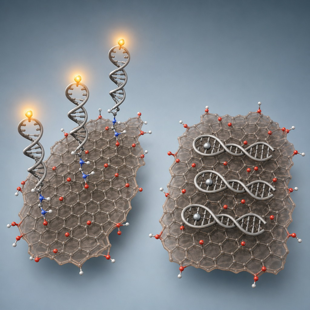
Carboxyl GO from ACS Material (Medford, MA) was the central material in every sensor variant. The authors selected this product because GO edges are naturally rich in carboxyl groups that can be activated by EDC for amide coupling to amino-terminated DNA, and because their dynamic light scattering measurements confirmed flake sizes greater than 2 µm, large enough to be reliably pelleted by centrifugation during the multi-step washing protocol. For covalent sensors, 200 µg/mL GO was reacted overnight with 4 µM amino-modified hairpin DNA in MES buffer with 10 mM EDC·HCl. Physisorbed DNA was then stripped using sequential 8 M urea washes, 80% isopropanol, complementary-DNA displacement, and elevated-temperature treatment to ensure that residual signal came only from covalently bound probes. Parallel physisorbed sensors used the same DNA and the same GO without EDC activation. Each probe carried an internal FAM fluorophore positioned at the hairpin loop so that Hg2+-induced folding would lift the dye 5, 9, or 13 base pairs away from the GO surface.
The coupling efficiency for covalent attachment was estimated at approximately 20% based on comparison with free-DNA fluorescence and a previously measured quenching efficiency of ~90% for duplex DNA on GO. Upon adding 1 µM Hg2+, both sensor types responded, but with characteristic differences. Physisorbed sensors gave larger absolute signal swings because Hg2+ binding fully desorbs the DNA from GO. Covalent sensors gave smaller but cleaner responses with markedly better selectivity against competing metal ions including Ce3+, Mn2+, Mg2+, Co2+, Cu2+, Zn2+, Cd2+, Pb2+, Ca2+, and Ag+. Critically, when BSA, Tween 80, or Triton X-100 (each at 0.1%) was added, physisorbed probes were displaced and produced large false-positive signals, while covalent sensors remained stable. The covalent sensor was also reversible: sequential additions of Hg2+ and KI (which sequesters Hg2+ as HgI4 2-) cycled the fluorescence signal up and down repeatedly, whereas the physisorbed sensor could not be regenerated because the desorbed DNA does not re-adsorb under sensing conditions. Probe 3, with 10 thymine-thymine Hg2+ binding sites, gave the strongest discrimination. Gel-based PAGE assays confirmed that Hg2+ released DNA into the supernatant only for the physisorbed system.
The results have clear implications for designing GO-based biosensors that must operate in complex biological or environmental matrices. Covalent DNA-GO conjugates are the preferred architecture whenever surfactants, proteins, or repeated sensor reuse are involved, including continuous water-monitoring devices, point-of-care diagnostic strips, and reusable lab-on-chip mercury sensors. Physisorbed sensors remain attractive for single-shot assays in clean buffer where signal magnitude matters most. The same design principles extend to aptamer-based detection of small molecules, protein biomarkers, and other heavy metals such as Pb2+ via DNAzyme chemistry. The authors specifically point to reusable and integrated biosensing devices as the most promising follow-up direction, where the demonstrated reversibility of covalent DNA-GO sensors becomes a decisive advantage.
For researchers exploring nucleic acid biosensors, electrochemical DNA platforms, or functionalized graphene chemistry, the carboxyl graphene oxide used in this study is available from ACS Material along with a broader range of oxidized and functionalized graphenes. The paper is a useful reference for anyone deciding between covalent and physisorbed probe architectures: it shows that the right choice depends on the sample matrix and the need for reusability, not on a universal preference for one strategy.How ACS Material products were used
- Carboxyl Graphene Oxide (carboxyl GO) (Graphene Series) — “Carboxyl GO was purchased from ACS Material (Medford, MA).”
Product Performance in this StudyCarboxyl-functionalized graphene oxide from ACS Material served as the central fluorescence-quenching scaffold for both covalently linked and physisorbed DNA biosensors. Its edge carboxyl groups enabled EDC-mediated amide coupling with amino-modified DNA probes, while its 2D surface efficiently quenched fluorophores until Hg2+-induced hairpin formation or desorption restored emission.
Related product categories
Frequently asked questionsWhy use covalent DNA attachment instead of physisorption on graphene oxide for biosensing?
Covalent attachment via EDC coupling produces sensors that resist displacement by surfactants and proteins, maintain selectivity against competing metal ions, and can be regenerated for repeated use. Physisorbed sensors give larger initial signals because target binding fully desorbs the DNA, but they cannot be reused and produce false positives in samples containing BSA, Tween 80, or Triton X-100 at concentrations as low as 0.1%.
How does carboxyl graphene oxide enable DNA covalent immobilization?
Carboxyl graphene oxide carries carboxylic acid groups on its edges that can be activated by EDC (1-ethyl-3-(3-dimethylaminopropyl)carbodiimide) to react with amino-terminated DNA, forming stable amide bonds. In this study, 200 µg/mL carboxyl GO from ACS Material reacted overnight with 4 µM amino-DNA in MES buffer at pH 6.0, achieving approximately 20% coupling efficiency after rigorous washing with urea, isopropanol, and complementary DNA.
What makes T-rich DNA probes selective for mercury ion detection?
Thymine bases form a Hg2+-mediated T-T base pair that is thermodynamically more stable than a normal T-A pair. When a thymine-rich hairpin DNA encounters Hg2+, it folds into a stable duplex through these T-Hg2+-T linkages, displacing the fluorophore away from a quenching surface like graphene oxide. Probes with more T-T binding sites (10 in this study's best probe) give stronger and more selective signal changes.