-
Carboxylated Graphene Quantum Dots for Photothermal Therapy - Università Cattolica, 2022
Jun 26, 2026 | ACS MATERIAL LLCPerini, G. et al. (2022). INSIDIA 2.0 high-throughput analysis of 3D cancer models: multiparametric quantification of graphene quantum dots photothermal therapy for glioblastoma …. *International Journal of Molecular Sciences*. https://doi.org/10.3390/ijms23063217
Dipartimento di Neuroscienze, Università Cattolica del Sacro Cuore, Largo Francesco Vito 1, 00168 Rome, Italy · International Journal of Molecular Sciences · 2022
Università Cattolica researchers used ACS Material carboxylated graphene quantum dots for NIR photothermal therapy of glioblastoma and pancreatic cancer spheroids, quantified with INSIDIA 2.0.
About this research
Researchers at Università Cattolica del Sacro Cuore used carboxylated graphene quantum dots (GQDs) purchased from ACS Material to deliver near-infrared photothermal therapy (PTT) against three-dimensional glioblastoma and pancreatic cancer spheroids, reducing spheroid volume by up to 70% over 14 days when combined with chemotherapy. The study introduces INSIDIA 2.0, an open-source ImageJ/FIJI macro that performs high-throughput, multiparametric quantification of spheroid morphology and grayscale texture. By coupling the GQD-based PTT with this software, the team distinguished two mechanistically different modes of spheroid death and built a single quantitative "spheroid disruption" parameter to score therapeutic efficacy non-invasively.
This research matters because chemotherapeutics often carry severe systemic toxicity and limited tumor penetration, motivating nanoparticle-based targeted therapies. Graphene quantum dots are attractive in oncology because they show high biocompatibility, low cytotoxicity, the ability to cross biological barriers, and rapid renal clearance owing to their small size. Critically, GQDs strongly absorb NIR light and convert it into localized heat, enabling photothermal ablation of tumor cells. At the same time, the rise of 3D spheroid models for drug screening has outpaced the tools available to analyze them: most current methods rely on manual measurement, are slow, and vary between operators. The combination of an efficient photothermal nanomaterial with an automated, customizable analysis pipeline addresses both the therapeutic and the screening bottlenecks faced by nanomedicine and cancer drug-discovery laboratories.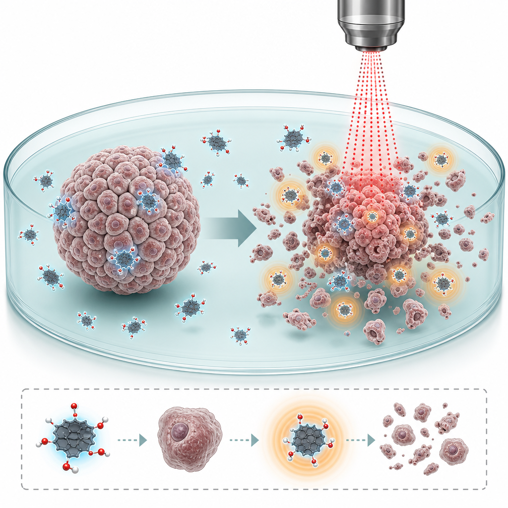
The carboxylated GQDs were supplied by ACS Material as a 1 mg/mL aqueous (ddH2O) dispersion. The team characterized the material before use: dynamic light scattering gave a hydrodynamic radius below 10 nm, atomic force microscopy on mica confirmed a maximum topographic height of about 0.8 nm, and fluorescence spectroscopy showed a peak emission at 450 nm (blue) under 320 nm excitation. ATR-FTIR confirmed the surface carboxyl chemistry, showing O–H stretches near 3000–2800 cm⁻¹ and a clear C=O stretch at 1750 cm⁻¹. For therapy, spheroids were treated with GQDs at 200 µg/mL for two weeks, with medium refreshed every three days, with or without doxorubicin (1 µM, U87) or 5-fluorouracil (100 µM, PANC-1). Photothermal activation used an 808 nm laser at 6 W/cm² for 5 min over a 0.8 cm spot, with a thermal camera monitoring temperature. In culture medium, the GQDs reached 42 °C within 5 min while the medium-only control showed no rise, confirming the photothermal conversion that drives the therapy.
The quantitative results show clear, model-specific responses. In U87 glioblastoma spheroids, untreated controls grew to about 150% of their initial area over 14 days, whereas 200 µg/mL GQDs alone halted growth. Doxorubicin immediately blocked growth and shrank spheroids by roughly 30% after 5 days, similar to Dox plus GQDs; however, adding PTT to the doxorubicin–GQD combination reduced spheroid volume to 70% of the initial value after 14 days. Area measurements correlated strongly with viability (R = 0.929). INSIDIA 2.0 also detected decreased circularity, increased specific surface, reduced core area, and lower grayscale standard deviation and GLCM entropy, indicating death by collapse into a uniform high-density core. In PANC-1 pancreatic spheroids, 5-fluorouracil alone cut viability by 20% and 70% after 7 and 14 days; combining 5FU with GQDs or GQD-PTT enhanced the drug effect and caused complete loss of spheroid integrity by day 14. Here, total area paradoxically increased while viability fell, with rising entropy and shrinking core area reflecting disaggregation. The authors captured both behaviors in a spheroid disruption parameter that becomes negative when area and core percentage diverge, flagging a death process.
The work enables faster, lower-cost, non-invasive screening of nanoparticle and combination therapies on 3D tumor models, relevant to precision nanomedicine, drug discovery, and photothermal oncology. Because INSIDIA 2.0 is open source, customizable, and compatible with other segmentation tools and imaging platforms, it can remove non-efficacious nanoparticle candidates earlier in development pipelines. The authors point to future studies using spheroids grown within extracellular matrix to clarify the biological significance of the distinct death modes, and they note the macro's versatility extends beyond oncology into materials science. The GQD-PTT strategy itself supports continued exploration of NIR-responsive carbon nanomaterials for treating hard-to-reach tumors such as glioblastoma.
For researchers working on photothermal therapy, biosensing, or nanoparticle-drug combinations, the carboxylated graphene quantum dots used here are available from ACS Material's quantum dots product line. The paper's characterization data — sub-10 nm hydrodynamic size, ~0.8 nm height, blue emission, defined carboxyl surface chemistry, and reproducible photothermal heating to 42 °C — provide a useful benchmark for groups planning similar 3D spheroid screening or NIR phototherapy experiments. As always, the material's value should be judged by the quantitative outcomes reported rather than by promotional claims.How ACS Material products were used
- Carboxylated Graphene Quantum Dots (Quantum Dots & Upconverting Nanoparticles) — “Carboxylated GQDs in double-distilled water (ddH2O) solution with a concentration of 1 mg/mL have been purchased from ACS Material (Pasadena, CA, USA).”
Product Performance in this StudyThe carboxylated GQDs absorbed 808 nm NIR light and converted it to heat, reaching 42 °C within 5 min, enabling effective photothermal disruption of glioblastoma and pancreatic cancer spheroids when combined with chemotherapy.
Related product categories
Frequently asked questionsHow do graphene quantum dots enable photothermal cancer therapy?
Graphene quantum dots strongly absorb near-infrared light and convert it into localized heat. In this study, carboxylated GQDs dispersed in culture medium reached 42 °C within 5 minutes under an 808 nm laser at 6 W/cm², while medium alone showed no temperature rise. This photothermal heating disrupted glioblastoma and pancreatic cancer spheroids, especially when combined with chemotherapy.
What concentration of graphene quantum dots was used to treat cancer spheroids?
The researchers treated U87 glioblastoma and PANC-1 pancreatic cancer spheroids with carboxylated graphene quantum dots at 200 µg/mL for two weeks, refreshing the medium with the same nanoparticle concentration every three days. The GQDs were supplied as a 1 mg/mL aqueous stock and diluted for treatment, optionally combined with doxorubicin or 5-fluorouracil.
Why is multiparametric image analysis important for screening nanoparticle therapies on 3D tumor models?
Cancer cells can die in different ways: glioblastoma spheroids collapsed into a uniform dense core with decreasing area and entropy, while pancreatic spheroids disaggregated with increasing area and entropy. A single metric like spheroid size can be misleading, since area may rise even as viability falls. Multiparametric analysis with INSIDIA 2.0 captures these distinct behaviors non-invasively and quantifies therapy efficacy reliably.