-
CVD Graphene FET for DNA Nucleobase Sensing - University of Melbourne, 2015
May 28, 2026 | ACS MATERIAL LLCDontschuk, N. et al. (2015). A graphene field-effect transistor as a molecule-specific probe of DNA nucleobases. *Nature Communications*. https://doi.org/10.1038/ncomms7563
Nature Communications · 2015
University of Melbourne researchers used CVD graphene from ACS Material to build field-effect transistors that detect molecule-specific DNA nucleobase signals.
About this research
Researchers at the University of Melbourne, in collaboration with La Trobe University and the Australian Synchrotron, used CVD graphene on copper foil sourced from ACS Material LLC to construct graphene field-effect transistors (GFETs) that distinguish the four DNA nucleobases by their adsorption-induced electronic signature. Published in Nature Communications in 2015, the work demonstrates that adenine, guanine, cytosine, and thymine each produce a distinct, coverage-dependent shift in the graphene charge neutrality point (CNP), which the authors attribute to interfacial dipole field formation rather than direct charge transfer. The result provides the first experimental electrical-transport evidence of molecule-specific nucleobase–graphene interactions, a long-standing assumption underpinning graphene-based DNA sequencing proposals.
Why this research matters: fast, label-free DNA sequencing remains a central biomedical goal, and graphene's two-dimensional structure, exceptional sensitivity, and low intrinsic noise make it a leading candidate for single-molecule electrical readout in nanopore and nanoribbon devices. Theoretical density functional theory studies had predicted distinguishable signatures from the four bases, but until this study no electrical transport experiment had confirmed that GFETs can resolve different nucleobases purely through physisorption. Establishing this experimentally addresses a fundamental open question: whether graphene, without chemical functionalization, retains enough analyte specificity to underpin sequencing technologies. The answer matters not only for sequencing but also for broader chemical and biological sensing platforms based on 2D materials.
How the ACS Material product was used: the authors fabricated GFETs from single-layer and bilayer CVD graphene grown on copper and transferred to 90 nm SiO2/Si substrates, obtained commercially from two suppliers, Graphene Laboratories Inc. and ACS Materials LLC. Two suppliers were deliberately used to test whether intrinsic defects or impurities in any single batch could bias the results. Channels were patterned 50 µm wide and 50–200 µm long using PMMA and oxygen plasma, with shadow-mask-evaporated gold contacts. Samples were rinsed in acetone, annealed at 250 °C in pure argon for 48 hours, and further annealed in ultra-high vacuum until the CNP stabilized. Large adjacent areas of the same ACS Material graphene were left contact-free for synchrotron X-ray photoelectron spectroscopy (XPS) and near-edge X-ray absorption fine structure (NEXAFS) measurements on the Soft X-ray beamline at the Australian Synchrotron. DNA nucleobase powders were sublimed in situ from low-temperature effusion cells at ~10⁻⁹ mbar onto the cleaned graphene surface.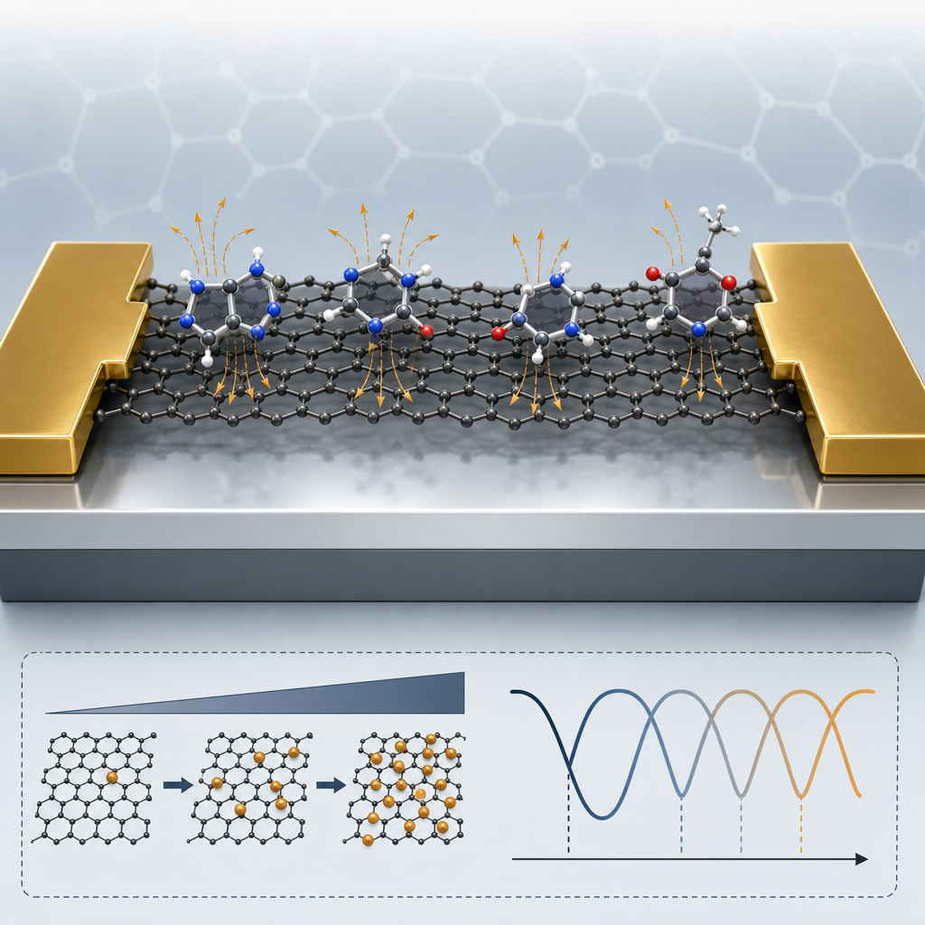
Key results: a 0.1 monolayer (~10⁹ molecules) dose of guanine shifted the CNP by 0.7 V, equivalent to ~1.7 × 10¹¹ additional electrons cm⁻². Across all four nucleobases the induced charge carrier density scaled linearly with coverage at low coverages and saturated at higher coverages, consistent with an electrostatic depolarization model in which mutual dipole–dipole interactions suppress the per-molecule contribution. The ordering of induced shifts at low coverage was G > C > T > A, matching the known order of nucleobase dipole moments. XPS C 1s binding-energy shifts measured simultaneously on identically prepared graphene gave the same qualitative ordering, with magnitudes up to ~5× larger than the CNP-derived Fermi-level shifts, indicating that the Ti/Au contacts limit GFET sensitivity. NEXAFS revealed a ~30–45° tilt of the nucleobase rings relative to the graphene plane and no π* hybridization, confirming pure van der Waals physisorption. Extrapolating the low-coverage linear regime to a 100 × 100 nm² device predicts single-molecule signals of Δn ≈ 10⁶–10⁸ cm⁻² per nucleobase, with guanine giving the largest response of 3.8 × 10⁸ cm⁻². With reported mobility and 1/f-noise improvements, single-molecule detection of guanine, cytosine, and thymine appears achievable.
Applications and outlook: the findings strengthen the case for graphene-based label-free DNA sequencing, particularly architectures combining nanopores or sub-nanometer armchair-edge nanoribbons with GFET readout. Beyond sequencing, the demonstrated ability of pristine graphene to resolve weakly interacting molecules by their dipole signatures is directly relevant to gas sensing, biomarker detection, and interface-dipole engineering of organic/2D heterostructures. The authors highlight three engineering levers for future work: improved metal–graphene contact alignment, substrate selection to suppress water-induced doping, and channel scaling into the nanoribbon regime where edge states may further enhance selectivity. Buffered-solution operation and discrimination against the charged phosphate backbone remain open challenges.
Why this matters for researchers: the study confirms that commercially available CVD graphene on copper foil, including material from ACS Material LLC, is suitable as the active channel for sensitive biosensing devices when paired with rigorous cleaning, UHV characterization, and contact engineering. Groups developing GFET biosensors, nanopore sequencing platforms, or 2D-material chemical sensors can source equivalent CVD graphene from ACS Material's CVD graphene catalog to reproduce and extend this work, as well as related transfer films and SiO2/Si substrates used throughout the GFET fabrication workflow.How ACS Material products were used
- CVD Graphene on Copper Foil (transferred to SiO2/Si) (CVD Graphene) — “SLG and BLG grown by CVD on Cu and transferred to SiO2 (90 nm)/Si substrates were obtained commercially from Graphene Laboratories Inc and ACS Materials LLC.”
Product Performance in this StudyThe CVD graphene served as the active channel of the field-effect transistor. Devices fabricated from ACS Material's CVD graphene reproduced the same nucleobase-induced conductance signatures as samples from the alternate supplier, confirming the sensing behavior was intrinsic to bulk CVD graphene rather than a unique defect or contaminant.
Related product categories
Frequently asked questionsHow does CVD graphene detect different DNA nucleobases in a field-effect transistor?
When adenine, guanine, cytosine, or thymine physisorb onto the graphene channel, each molecule forms an interfacial dipole that shifts the graphene charge neutrality point. The shift scales linearly with coverage at low doses and saturates at high coverage as dipole–dipole depolarization sets in. Because each nucleobase has a distinct dipole moment, each produces a characteristic conductance signature without any chemical functionalization of the graphene surface.
Why is CVD graphene on copper foil preferred for GFET biosensing channels?
CVD graphene grown on copper and transferred to SiO2/Si offers large-area, single-layer films compatible with photolithography and shadow-mask contact deposition. In this study, channels several times larger than the typical 5–10 µm² grain size were used to average out grain-boundary effects and probe intrinsic bulk transport. Using CVD graphene from two suppliers, including ACS Material, also let the authors confirm the nucleobase response was reproducible rather than supplier-specific.
What sensitivity can graphene FETs achieve for single-molecule DNA detection?
Extrapolating the measured low-coverage response to a 100 × 100 nm² channel predicts single-molecule signals of roughly 10⁶–10⁸ cm⁻² induced carriers, with guanine producing the largest shift of 3.8 × 10⁸ cm⁻². Current device noise is around 10¹⁰ cm⁻², but the authors estimate that combining high-mobility graphene (up to 2 × 10⁵ cm² V⁻¹ s⁻¹), optimized contacts, and reduced 1/f noise could yield single-molecule resolution for guanine, cytosine, and thymine.