-
Graphene Oxide DNA Sensing Mechanism - University of Waterloo, 2013
Jun 19, 2026 | ACS MATERIAL LLCLiu, B. et al. (2013). Mechanisms of DNA sensing on graphene oxide. *Analytical Chemistry*. https://doi.org/10.1021/ac401845p
University of Waterloo · Analytical Chemistry · 2013
University of Waterloo researchers used ACS Material graphene oxide to reveal that DNA sensing on GO follows a displacement-then-hybridization mechanism.
About this research
Researchers at the University of Waterloo used graphene oxide (GO) purchased from ACS Material to resolve a long-standing question about how fluorescent DNA biosensors actually work on a GO surface, demonstrating that hybridization proceeds through a non-specific displacement step followed by duplex formation in solution rather than on the nanosheet itself. The team, led by Juewen Liu, designed a series of kinetic and thermal-desorption experiments using fluorophore-labeled poly-A and poly-T probes to rule out the long-assumed Langmuir-Hinshelwood and Eley-Rideal mechanisms. Their conclusion reframes how the field should approach rational sensor design and signal optimization in GO/DNA systems.
GO-based fluorescence biosensors have become a standard tool for detecting nucleic acids, metal ions, small molecules, proteins, and even whole cells, valued for their simplicity, high signal-to-background ratio, and sub-nanomolar sensitivity. The platform exploits two complementary properties of graphene oxide: strong adsorption of single-stranded DNA through π–π stacking, hydrophobic interactions, hydrogen bonding, and van der Waals forces; and efficient fluorescence quenching of bound fluorophores. Despite hundreds of analytical demonstrations, the molecular-level mechanism by which a target DNA triggers turn-on fluorescence had not been experimentally established. Most prior physical studies measured only adsorption energies and lacked dynamic information, so it was unclear whether hybridization occurred directly on the GO surface or in the surrounding solution. Resolving this question is essential not only for biosensing but also for related GO applications in targeted drug delivery, imaging, and bio-nano interface engineering.
Graphene oxide from ACS Material was the central material throughout this study. The authors dispersed GO in HEPES buffer with MgCl2 and adsorbed FAM-labeled, Alexa Fluor 647-labeled, or dual-labeled DNA probes at concentrations between 5 nM and 2 µM on 50 µg/mL GO. After incubation, free DNA was removed by repeated centrifugation (15,000 rpm, 20 min) and washing. The DNA/GO complexes were then exposed to complementary or non-complementary DNA targets while fluorescence kinetics were tracked on a plate reader or a real-time PCR thermocycler. Thermal desorption profiles, dual-fluorophore quantification of adsorbed versus desorbed DNA populations, and hybridization-rate comparisons with and without GO were all performed on this single GO source. A TEM micrograph of the as-received GO is included in the Supporting Information.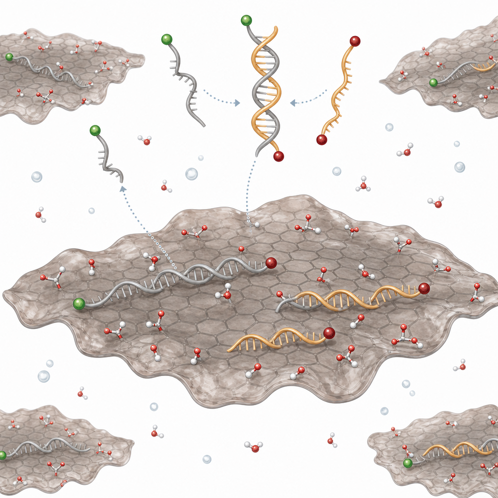
Key results center on the asymmetric behavior of poly-A and poly-T probes, chosen to maximize adsorption-energy contrast (purines bind GO more strongly than pyrimidines). FAM-A15 showed almost no thermal desorption from GO, while FAM-T15 desorbed with a melting temperature of ~60 °C, compared with ~44 °C for the free T15/A15 duplex—indicating that ss-DNA on GO is more stable than the free duplex. Adding 200 nM cDNA to pre-adsorbed FAM-A15 gave only a slow, weak signal, ruling out the Eley-Rideal mechanism. Adsorption kinetics for both A15 and T15 were complete within ~20 s, much faster than desorption, so differences in signal kinetics could not be explained by Langmuir-Hinshelwood surface diffusion. Dual-label experiments quantified the partition: when AF-T15 was the probe and 200 nM FAM-A15 was added, ~14 nM duplex formed, ~80 nM FAM-A15 adsorbed onto GO, and ~106 nM remained in solution. Only ~15% of cDNA contributed to signal—about six to seven equivalents of target are required to displace one adsorbed probe. Hairpin-probe controls further showed that GO actually inhibits hybridization rather than catalyzing it, disproving any catalytic surface mechanism.
These mechanistic insights have direct implications for biosensor engineering. Knowing that signal generation is gated by non-specific displacement rather than on-surface hybridization, researchers can now target two clear levers for improving sensitivity: blocking non-specific adsorption sites on GO after probe loading, and deliberately tuning the adsorption-energy gap between probe and target. The findings extend to aptamer-based sensors for metal ions, small molecules, proteins, and cells, and inform GO use in targeted drug delivery, fluorescent imaging probes, and broader 2D-material bio-interfaces. The displacement framework can also guide the design of molecular beacons, strand-displacement amplification on nanosheets, and competitive assays on other 2D platforms such as MoS2, WS2, and hexagonal boron nitride.
For researchers working on GO-based biosensors, drug delivery vehicles, or interfacial DNA studies, the consistent, well-characterized graphene oxide supplied by ACS Material was central to obtaining reproducible kinetics across thermal desorption, dual-fluorophore tracking, and hairpin-probe experiments. The graphene oxide and related graphene-series materials used here remain available for laboratories developing turn-on fluorescent assays, aptasensor platforms, and 2D-material bio-interfaces. This paper offers a reference framework for any group designing GO/DNA experiments where mechanistic clarity matters as much as raw sensitivity.How ACS Material products were used
- Graphene Oxide (GO) (Graphene Series) — “The GO sample was purchased from Advanced Chemical Supplier (ACS) Material (Medford, MA).”
Product Performance in this StudyThe ACS Material graphene oxide served as the active surface for fluorophore-labeled DNA adsorption and the central platform on which the displacement-then-hybridization sensing mechanism was elucidated. It enabled strong quenching, low background fluorescence, and reproducible adsorption/desorption kinetics suitable for mechanistic study.
Related product categories
Frequently asked questionsHow does DNA hybridization actually occur on a graphene oxide biosensor?
Hybridization does not happen on the graphene oxide surface itself. Instead, added target DNA non-specifically competes with the adsorbed fluorophore-labeled probe for binding sites on GO. Some probe DNA is displaced into solution, where it then hybridizes with free target DNA to form a duplex. Because double-stranded DNA binds GO weakly, the duplex stays in solution and fluorescence is recovered, producing the turn-on signal.
Why is graphene oxide an effective platform for fluorescent DNA sensing?
Graphene oxide combines two useful properties: it adsorbs single-stranded DNA strongly through π–π stacking, hydrophobic forces, hydrogen bonding, and van der Waals interactions, and it efficiently quenches nearby fluorophores. This produces an extremely dark background when the probe is bound, so any release of probe DNA into solution yields a high signal-to-background ratio. Sensitivities down to about 1 nM DNA are routinely achieved despite low target-utilization efficiency.
How can researchers improve the efficiency of graphene oxide DNA biosensors?
Because only about 15 percent of added target DNA generates signal in optimized conditions, sensitivity gains require addressing the displacement bottleneck. Strategies include blocking non-specific adsorption sites on the graphene oxide surface after probe loading, designing probes with deliberately lower GO affinity than the target, and tuning nucleotide composition since purines bind GO more strongly than pyrimidines. Hairpin and molecular beacon probe geometries can further reduce background.