-
Graphene Oxide for Selective Lysozyme Adsorption — Cerritos College, 2014
May 26, 2026 | ACS MATERIAL LLCLi, S. et al. (2014). Strong and selective adsorption of lysozyme on graphene oxide. https://doi.org/10.1021/am500254e
2014
Cerritos College researchers used single-layer graphene oxide from ACS Material to selectively adsorb lysozyme from protein mixtures via electrostatic binding.
About this research
Researchers affiliated with Cerritos College, working with collaborators at the University of Miami and MP Biomedicals, used single-layer graphene oxide (GO) purchased from ACS Material LLC to investigate and quantify the strong, selective adsorption of lysozyme onto GO surfaces. Published in ACS Applied Materials & Interfaces in 2014, the study shows that the GO–lysozyme interaction is strong enough to fully sequester lysozyme from aqueous solution, to selectively capture it from binary and ternary protein mixtures, and to permit subsequent desorption under controlled pH and salt conditions. The work directly informs the design of GO-based diagnostic platforms for biological fluids.
This research matters because graphene oxide is increasingly used in biosensing, controlled drug delivery, cellular imaging, and photothermal therapy. For any diagnostic technology applied to real biological fluids — saliva, tears, milk, serum, urine — the sensor must reject interference from abundant background proteins. Lysozyme is present at roughly 21.4 μg/mL in human milk, 7 μg/mL in saliva, and as much as 1568 μg/mL in tears, and is dramatically elevated in patients with leukemia, renal disease, or sarcoidosis. If lysozyme adsorbs strongly and nonspecifically onto GO, it could mask intended analytes. Before this study, the nature of the GO–lysozyme interaction had not been systematically defined, leaving a gap in the protein-corona understanding required for translational GO biosensors.
The ACS Material single-layer GO was used as the active adsorbent throughout the study. A 1 mg/mL aqueous dispersion was prepared by dissolving 10 mg of GO in 10 mL of deionized water followed by one hour of bath sonication, then diluted with water or 0.1 M phosphate buffer (pH 7) as needed. For fluorescence quenching experiments, GO was titrated from 0 to 10 μg/mL against 1 × 10⁻⁶ M solutions of lysozyme, BSA, HSA, and ovalbumin. For adsorption experiments, 0.1 mg/mL GO was combined with 0.143 mg/mL lysozyme, with NaCl added to drive precipitation and centrifugation at 2500 rpm used to separate the protein-loaded GO. For desorption, the GO–lysozyme pellet was treated with pH 11.5 NaOH and sonicated, then GO was precipitated with 0.1 M CaCl₂ to leave released lysozyme in the supernatant. The same protocol with phosphate buffer was used to test selective adsorption from binary and ternary protein mixtures.
The results demonstrate that GO interacts far more strongly with lysozyme than with bovine serum albumin (BSA), human serum albumin (HSA), or ovalbumin (OVA). Fluorescence quenching of lysozyme by GO was substantially larger than for the other proteins after correction for the inner-filter effect, consistent with a dominant electrostatic interaction between negatively charged GO and positively charged lysozyme (lysozyme has a high isoelectric point near 11). Zeta-potential measurements at pH 5.6 confirmed charge-pairing: GO carried a strongly negative zeta potential while lysozyme carried a positive one, and the GO/LYZ mixture exhibited a zeta-potential trend tracking the lysozyme loading from 0 to 100 μg/mL. Dynamic light scattering and AFM showed that lysozyme binding produced GO sheets decorated with discrete protein features. At pH 5.6, GO completely removed lysozyme from solution; the protein could then be released by switching to pH 11.5 NaOH, which neutralizes the electrostatic attraction, followed by CaCl₂-induced GO precipitation. SDS-PAGE of binary mixtures (LYZ/BSA, LYZ/HSA, LYZ/OVA) and ternary mixtures (LYZ/OVA/BSA, LYZ/OVA/HSA) confirmed that only the lysozyme band was depleted from the supernatant after GO treatment, with the other proteins remaining in solution. Fluorescence and UV–vis spectra of the supernatants corroborated this selectivity.
These findings have direct implications for GO-based diagnostics intended for tears, milk, saliva, serum, or urine, where lysozyme is naturally abundant and clinically variable. The strong, pH-tunable, electrostatic capture of lysozyme suggests two parallel application paths: first, controlling or blocking the lysozyme corona before deploying GO sensors for other biomolecules; second, exploiting GO as a selective preconcentration platform for lysozyme itself in clinical samples relevant to leukemia, renal disease, and sarcoidosis screening. The reversible adsorption-desorption protocol — using NaCl-assisted precipitation and pH 11.5 elution — also provides a template for protein recovery in proteomic workflows. The authors highlight further work on GO surface chemistry tuning and on probing related cationic proteins.
For researchers planning similar adsorption, protein-corona, or GO-biosensor experiments, single-layer graphene oxide of the grade used here is available from ACS Material's Graphene Series. The paper's quantitative protocols — GO concentration, sonication, buffer choice, NaCl-assisted precipitation, and CaCl₂ elution — are reproducible starting points for new investigations into selective biomolecule capture, GO surface modification, and diagnostic platform development.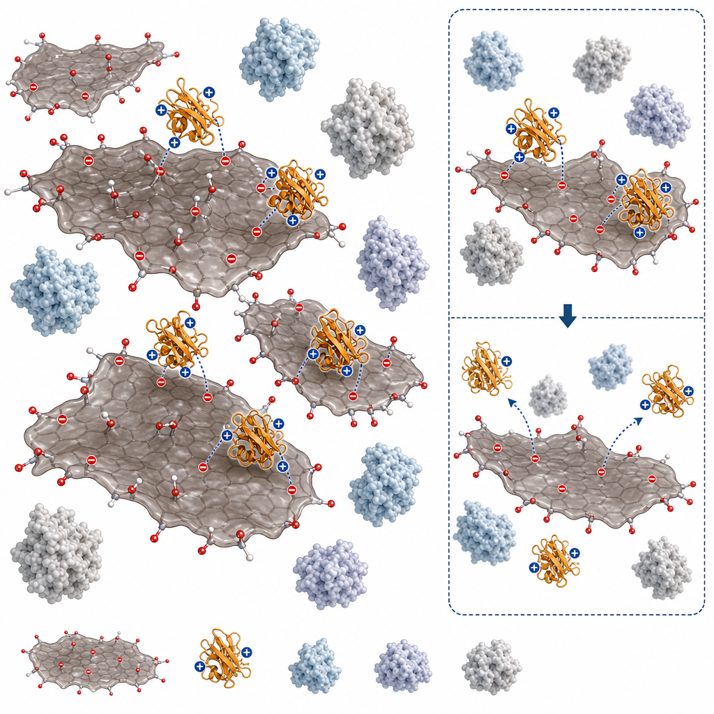
How ACS Material products were used
- Single-Layer Graphene Oxide (Graphene Series) — “Single-layer graphene oxide (GO) was bought from ACS Material LLC (Medford, MA).”
Product Performance in this Study
The single-layer graphene oxide from ACS Material served as the central adsorbent in the study, exhibiting strong electrostatic interaction and selective adsorption of lysozyme over BSA, HSA, and ovalbumin. The material's properties were consistent with prior characterizations and enabled both effective adsorption and subsequent desorption.
Related product categories
Frequently asked questions
Why does graphene oxide adsorb lysozyme more strongly than serum albumin?
The adsorption is driven primarily by electrostatic attraction. Graphene oxide carries a strongly negative surface charge from carboxyl, hydroxyl, and epoxide groups, while lysozyme has a high isoelectric point near 11 and remains positively charged at neutral pH. BSA, HSA, and ovalbumin have lower isoelectric points and become negative or near-neutral at pH 7, producing far weaker electrostatic affinity. This pairing yields strong fluorescence quenching and complete capture of lysozyme.
How can lysozyme be desorbed from graphene oxide after adsorption?
The authors release adsorbed lysozyme by raising the pH to 11.5 with NaOH and sonicating briefly. At this pH the electrostatic attraction between GO and lysozyme is neutralized because lysozyme approaches its isoelectric point and loses positive charge. Calcium chloride (0.1 M CaCl₂) is then added to precipitate the GO sheets, leaving free lysozyme in the supernatant for recovery or analysis.
Does graphene oxide selectivity for lysozyme work in mixed protein samples?
Yes. SDS-PAGE results from binary mixtures of LYZ with BSA, HSA, or OVA, and ternary mixtures (LYZ/OVA/BSA and LYZ/OVA/HSA), showed that GO depleted only the lysozyme band while the other proteins remained in solution. Fluorescence and UV–vis spectra of supernatants confirmed selective lysozyme removal, supporting GO as a selective adsorbent in complex biological matrices.