-
Graphene Oxide microRNA Immunosensor - Univ. Paris Diderot, 2013
Jun 19, 2026 | ACS MATERIAL LLCTran, H. V. et al. (2013). Antibodies directed to RNA/DNA hybrids: an electrochemical immunosensor for microRNAs detection using graphene-composite electrodes. *Analytical Chemistry*. https://doi.org/10.1021/ac402154z
Univ. Paris Diderot, Sorbonne Paris Cité, ITODYS, UMR 7086 CNRS, 15 rue J-A de Baïf, 75205 Paris Cedex 13, France · Analytical Chemistry · 2013
Reduced graphene oxide composite electrochemical biosensor detects miR-29b-1 and miR-141 at ~5 fM with On-Off-On antibody triple verification.
About this research
Researchers at Univ. Paris Diderot, Sorbonne Paris Cité, ITODYS, UMR 7086 CNRS, 15 rue J-A de Baïf, 75205 Paris Cedex 13, France used single-layer graphene oxide purchased from ACS Material LLC to build a label-free electrochemical microRNA immunosensor that detects miR-29b-1 (a myocardial infarction biomarker) and miR-141 (a prostate cancer biomarker) down to a quantification limit of about 5 fM. The team, led by M.C. Pham, combined hybridization-based signal-on detection with anti-RNA/DNA antibody complexation to deliver a triple On-Off-On verification sequence, increasing reliability over conventional electrochemical biosensors. The work appeared in Analytical Chemistry in 2013.
MicroRNAs are short, non-coding regulatory RNAs whose dysregulation is implicated in cancers, cardiovascular disease, and many other disorders. Established quantification methods such as Northern blot, microarray, and qRT-PCR are sensitive but expensive, slow, and unsuited to point-of-care multiplexing. Electrochemical biosensors offer a cheaper, faster route, and combining them with nanostructured carbon supports such as graphene markedly increases electroactive surface area and electron-transfer kinetics. Adding anti-RNA/DNA antibodies that recognize heteroduplex structures regardless of nucleotide sequence brings a second layer of selectivity on top of standard Watson–Crick hybridization. The challenge is reaching sub-picomolar detection in body fluids without enzymatic amplification while still discriminating single-base mismatches, which this paper addresses.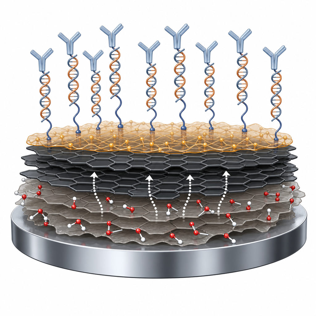
The ACS Material single-layer graphene oxide had a flake diameter of 1–5 μm and thickness of 0.8–1.2 nm, prepared by a modified Hummers method. Twenty-four milligrams were dispersed in water by ultrasonication, then reduced with epigallocatechin gallate (EGCG) at 80 °C for 8 h to give a stable aqueous reduced graphene oxide (RGO) suspension. Five microliters of 0.5 mg/mL RGO was drop-cast onto polished glassy carbon electrodes to give a surface density of 35.7 μg/cm². A conducting poly(JUG-co-JUGA) film was then electropolymerized on top from acetonitrile by potential cycling between 0.4 and 1.1 V vs SCE for 25 cycles. AFM showed RGO sheets ~2 nm thick on the electrode, slightly thicker than the starting GO due to EGCG intercalation, and UV–vis confirmed restoration of π-conjugation with a red shift from 234 to 269 nm. NH2-modified DNA probes were finally grafted via EDC/NHS chemistry to the conducting-polymer carboxyls, producing the CP/RGO/GCE biosensor platform.
Charge integration showed the RGO layer roughly tripled the electroactive quinone surface density of the conducting polymer, from (3.51 ± 0.48) × 10⁻⁹ mol/cm² on bare GCE to (10.57 ± 1.43) × 10⁻⁹ mol/cm² on RGO/GCE. Square-wave voltammetry gave a strong signal-on response upon miRNA hybridization, with current increasing monotonically over a target concentration range from 1 fM to 10 nM and a practical limit of quantification of ~5 fM. Melting-curve experiments on miR-29b-1/probe duplexes yielded an apparent melting temperature near 55 °C, consistent with the expected RNA/DNA duplex stability. Selectivity was strong: cross-hybridization tests with mismatched probes gave only minor current changes, and in a binary mixture the sensor distinguished 1 pM miR-29b-1 from miR-141 (21% vs 4% signal change). Two anti-RNA/DNA antibodies—polyclonal antipoly(A)–poly(dT) from Stollar's group and monoclonal S9.6 from Leppla's group—then complexed to the surface heteroduplex, producing a signal-off current decrease of ~25% for specific targets vs ≤2% for nonspecific anti-NEF controls, with an antibody-confirmed detection limit of ~8 fM. Adding free miRNA/DNA hybrids into solution competitively displaced the antibodies and returned the signal-on state, completing the On-Off-On cycle.
This sensor design opens a practical route to electrochemical microRNA biomarker assays for cancer and cardiovascular disease without PCR or enzymatic amplification. Because anti-RNA/DNA antibodies recognize hybrids in a sequence-independent manner, the same antibody reagent can validate any miRNA target once a complementary probe is grafted, simplifying multiplexed panel development. Adjacent application areas include point-of-care nucleic-acid diagnostics, liquid biopsy, circulating tumor marker quantification, and integration into screen-printed or microfluidic electrode arrays. The reduced graphene oxide composite electrode strategy also transfers readily to other DNA aptasensor or protein-binding formats where high surface area and good electron transfer are needed.
For researchers building similar electrochemical biosensors, single-layer graphene oxide of the type used in this work is available from ACS Material in the graphene oxide product line, including monolayer dispersions and powders suitable for chemical or thermal reduction to RGO. The paper's quantitative comparison of RGO-modified vs bare glassy carbon electrodes provides a useful baseline for anyone planning conducting polymer/graphene nanocomposite biosensors, and the reagent specifications (1–5 μm flake size, sub-1.2 nm thickness) give a clear starting point for reproducing the electrode fabrication protocol.How ACS Material products were used
- Single-Layer Graphene Oxide (diameter 1-5 μm, thickness 0.8-1.2 nm) (Graphene Series) — “Single-layer grapheme oxide (diameter 1−5 μm; thickness 0.8−1.2 nm) was purchased from ACS Material LLC (Medford, MA, USA) and synthesized using the modified Hummer's method.”
Product Performance in this StudyThe single-layer graphene oxide from ACS Material served as the precursor for the reduced graphene oxide (RGO) nanocomposite electrode coating. After reduction with EGCG, it produced RGO sheets ~2 nm thick that tripled the electroactive quinone surface density (from 3.51 to 10.57 nmol/cm²) on the conducting polymer electrode.
Related product categories
Frequently asked questionsWhy use reduced graphene oxide in an electrochemical microRNA biosensor?
Reduced graphene oxide provides a high-surface-area, electrically conductive layer that anchors a conducting polymer film and increases the density of electroactive groups available for signal transduction. In this work, RGO drop-cast onto glassy carbon electrodes raised the electroactive quinone surface density from 3.51 to 10.57 nmol/cm² compared to bare GCE, enabling femtomolar microRNA detection with square-wave voltammetry without enzymatic amplification.
How do anti-RNA/DNA antibodies improve microRNA detection selectivity?
Antibodies such as S9.6 and antipoly(A)–poly(dT) bind RNA/DNA heteroduplexes in a sequence-independent manner. When they bind microRNA-DNA hybrids on the electrode surface, steric hindrance slows ion exchange and produces a clear current decrease (signal-off). This adds an immunological selectivity layer on top of Watson–Crick hybridization, letting the sensor confirm true hybridization events and distinguish them from nonspecific protein adsorption, which produces a different signal pattern.
What detection limits did the graphene oxide-based microRNA sensor achieve?
The CP/RGO/GCE sensor reached a practical quantification limit of approximately 5 fM for miR-29b-1 and miR-141 via direct hybridization-induced signal-on response, with a linear range from 1 fM to 10 nM. With anti-RNA/DNA antibody complexation as a second verification step, the antibody-confirmed detection limit was about 8 fM. Single-base mismatches and noncomplementary microRNAs produced negligible current change.