-
Graphene-Sealed Liquid Cell TEM of Au Nanoparticles - FAU Erlangen-Nürnberg, 2019
Jul 09, 2026 | ACS MATERIAL LLCHutzler, A. et al. (2019). In Situ Liquid Cell TEM Studies on Etching and Growth Mechanisms of Gold Nanoparticles at a Solid–Liquid–Gas Interface. *Advanced Materials Interfaces*. https://doi.org/10.1002/admi.201901027
Electron Devices (LEB) Department of Electrical, Electronic and Communication Engineering Friedrich‐Alexander University Erlangen‐Nürnberg Cauerstraße 6 91058 Erlangen Germany · Advanced Materials Interfaces · 2019
FAU Erlangen-Nürnberg uses ACS Material few-layer graphene to seal microwell liquid cells and image gold nanoparticle etching and growth in situ by TEM.
About this research
Researchers at Friedrich-Alexander University Erlangen-Nürnberg (Electron Devices, LEB) used few-layer (6–8 layers) graphene purchased from ACS Material to seal graphene-supported microwell liquid cells (GSMLCs) and directly observe the etching and regrowth of gold nanoparticles at a solid–liquid–gas interface by in situ liquid cell transmission electron microscopy (LCTEM). Working with a 10 mmol L⁻¹ aqueous HAuCl₄ precursor, the team captured how a primary Au nanoplatelet dissolved in the vicinity of a radiolytically generated gas bubble and how new AuNPs subsequently nucleated and grew in the confined liquid film between the bubble and the cell membrane. The work was published in Advanced Materials Interfaces in 2019.
Understanding reaction mechanisms at solid–liquid–gas interfaces is critical for designing greener industrial processes, including cyanide-free leaching of noble metals using halide chemistry. While liquid cell TEM has matured into a powerful tool for tracking nanoscale dynamics, most prior studies have focused on simple solid–liquid interfaces. Three-phase boundaries, where gas bubbles meet liquid and a solid nanoparticle, remain poorly characterized despite their relevance to electrocatalysis, corrosion, and metal recovery. The Erlangen team specifically targeted this gap, exploring how a chlorine-rich gas bubble alters the dissolution pathway of gold and how the dissolved species redeposit nearby. Long-tail terms relevant here include 'in situ liquid cell TEM gold', 'radiolysis HAuCl4', 'three-phase interface nanoparticle etching', and 'graphene-sealed microwell liquid cell'.
The ACS Material graphene played a structurally essential role. According to the Experimental Section, 'a R2/2 Quantifoil TEM grid covered with a few-layer (6–8 layers) graphene sheet purchased from ACS Material was placed on top of the microwell containing membrane with the graphene-covered side facing the liquid-cell template'. The graphene adhered to the silicon nitride surface via van der Waals interaction, hermetically enclosing 0.5 µL droplets of the HAuCl₄ solution within the microwells without epoxy or external clamping. The number of layers was independently verified by reflectance spectroscopy on a specialized optical stack. Because the graphene window is only a few atomic layers thick, it preserves high spatial resolution at 300 kV in a Philips CM 30 (S)TEM operated at electron dose rates of 10⁴–10⁵ e⁻ nm⁻² s⁻¹, while remaining strong enough to contain the aqueous gold precursor against vacuum.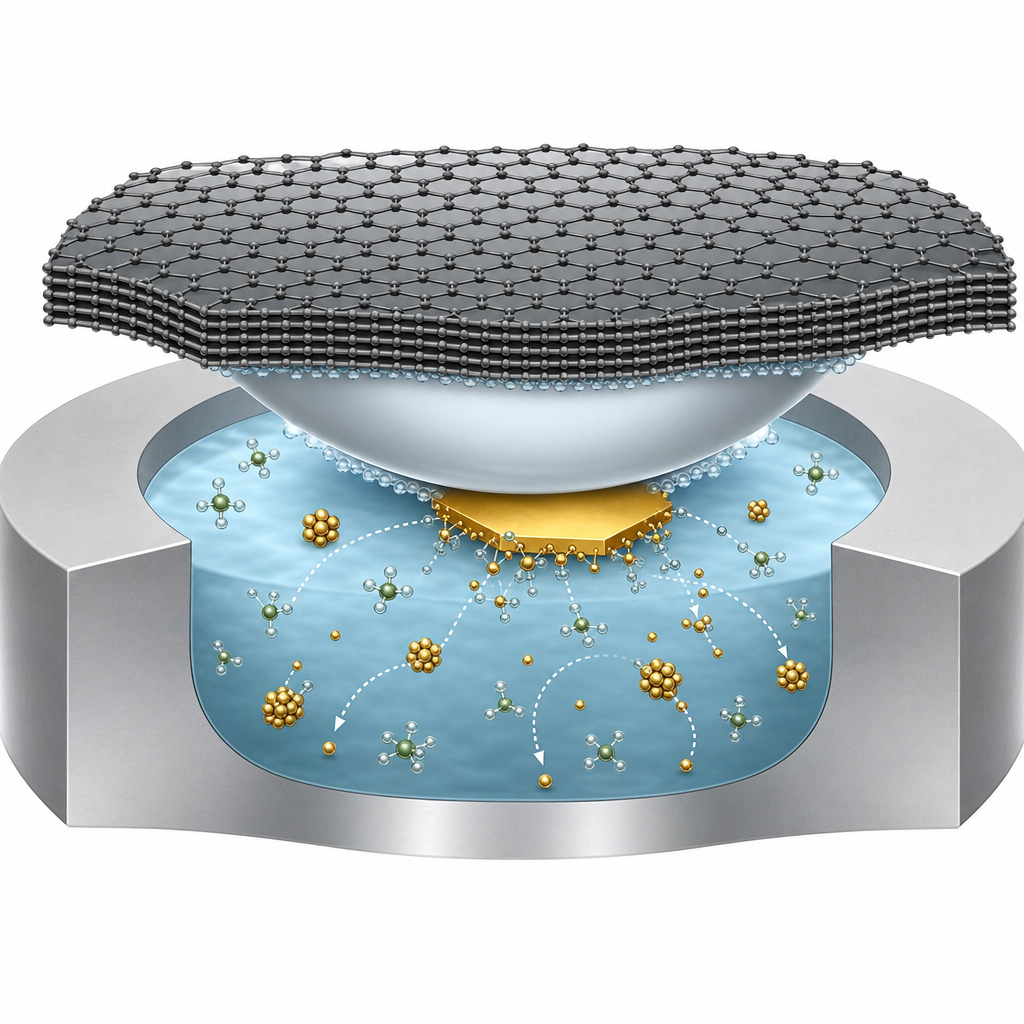
Key results centered on two regimes. Etching of the Au platelet proceeded markedly faster when the platelet contacted the gas bubble, separated only by a thin liquid wetting layer, than when it was fully immersed. The team extracted Wagner-type kinetic exponents for the equivalent particle radius: β evolved from −0.16 ± 0.04 to −1 ± 0.12 in the bubble-contact case (surface-reaction limited) and from −0.04 ± 0.01 to −1.41 ± 0.08 in the fully immersed case (diffusion limited). They attributed dissolution to OH• radicals generated by radiolysis oxidizing Au atoms, followed by complexation with Cl⁻ derived from gaseous Cl₂ and HCl trapped in the bubble. After the primary 9 nm platelet (≈1.44 nm thick) was consumed, the cumulative volume of newly nucleated AuNPs nearly matched the original platelet volume, indicating mass conservation within the confined volume. The growth exponent averaged 0.20 ± 0.15 across 50 particles—consistent with diffusion-limited growth (LSW predicts 1/3) but slowed by extreme confinement. By tracking particle trajectories and applying the 2D Stokes–Einstein relation, the authors determined the local dynamic viscosity to be 1.44 ± 0.04 MPa·s, dramatically higher than bulk water, reflecting narrow-channel effects in a liquid layer only a few nanometers thick. A critical nucleation radius near 0.5 nm was also identified.
These findings have practical implications for hydrometallurgy, electrocatalyst synthesis, and any process where gas evolution modifies metal dissolution—including chloride leaching of electronic waste and shape-controlled nanoparticle growth. The methodology also informs the design of in situ electrochemistry experiments, where bubble formation is often unavoidable. Adjacent application areas that benefit include plasmonic nanoparticle engineering, where AuNP shape and size dictate optical response, and corrosion science at gas-evolving electrodes. The authors point toward extending the approach to other halide-leaching chemistries and to bimetallic systems where selective dissolution drives morphology.
For researchers planning similar liquid cell TEM experiments, the few-layer graphene used here is offered by ACS Material as graphene on Quantifoil lacey carbon TEM grids and related transferable graphene products. The combination of mechanical robustness, electron transparency, and reliable van der Waals sealing to silicon nitride microwells makes these grids well suited to LCTEM workflows targeting nanoparticle dynamics, radiolysis-driven reactions, or three-phase interface studies. The performance reported in this paper—stable sealing over multi-minute imaging at 300 kV—is consistent with the product's specification as a thin, defect-tolerant support film.How ACS Material products were used
- Graphene on Lacey Carbon (Quantifoil R2/2) TEM Grids - few-layer (6-8 layers) graphene (CVD Graphene) — “A R2/2 Quantifoil TEM grid covered with a few-layer (6–8 layers) graphene sheet purchased from ACS Material was placed on top of the microwell containing membrane with the graphene-covered side facing the liquid-cell template.”
Product Performance in this Study
The ACS Material few-layer graphene sealed the microwell liquid cell via van der Waals adhesion to the silicon nitride membrane, providing an electron-transparent, leak-tight window that enabled atomic-scale in situ liquid cell TEM imaging of gold nanoparticle etching and growth at a solid–liquid–gas interface.
Related product categories
Frequently asked questions
Why use few-layer graphene to seal a liquid cell for TEM?
Few-layer graphene is only a few atomic layers thick, so it adds negligible scattering to the electron beam while preserving high spatial resolution at 300 kV. It also adheres to silicon nitride via van der Waals interaction, hermetically sealing aqueous samples in microwells without epoxy. In this study, 6–8 layer graphene from ACS Material allowed stable imaging of HAuCl4 solution and a gas bubble for many minutes under electron irradiation.
How does a gas bubble change the etching of gold nanoparticles in liquid cell TEM?
When a gas bubble directly contacts a gold nanoparticle, chlorine-containing gaseous species such as Cl2 and HCl permeate the thin liquid wetting layer and react at the particle surface, making etching surface-reaction limited and fast. When the particle is fully immersed, the same reactants must diffuse through bulk liquid, so etching becomes diffusion limited and slower. The measured Wagner exponents differed significantly between the two regimes.
Why was the measured viscosity inside the graphene liquid cell so high?
Using the 2D Stokes–Einstein relation and tracked particle trajectories, the authors obtained a dynamic viscosity of 1.44 ± 0.04 MPa·s, far above bulk water. This is attributed to narrow-channel effects: the liquid layer between the graphene-sealed silicon nitride membrane and the gas bubble is only a few nanometers thick, so interactions with both interfaces dominate and dramatically slow diffusion of solvated ions and nanoparticles.