-
Graphene–Gold Aptasensor for Norovirus Detection — University of Guelph, 2017
Jun 05, 2026 | ACS MATERIAL LLCChand, R., & Neethirajan, S. (2017). Microfluidic platform integrated with graphene-gold nano-composite aptasensor for one-step detection of norovirus. *Biosensors and Bioelectronics*.
Biosensors and Bioelectronics · 2017
University of Guelph researchers used ACS Material graphene dispersion to build a microfluidic graphene–gold aptasensor detecting norovirus down to 100 pM.
About this research
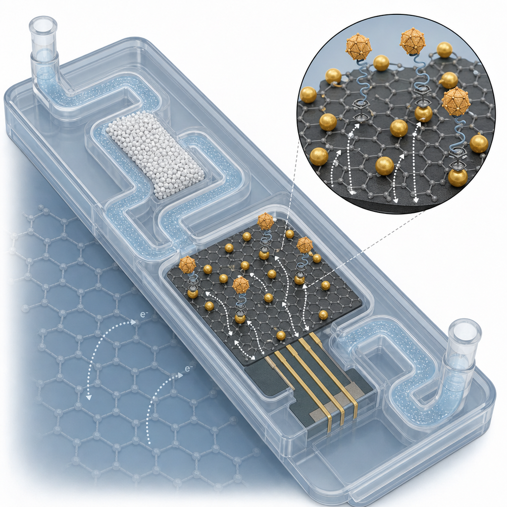
Researchers at the University of Guelph (Canada) used graphene dispersion purchased from ACS Material to fabricate a graphene–gold nanocomposite aptasensor integrated into a polydimethylsiloxane (PDMS) microfluidic chip, enabling one-step electrochemical detection of norovirus down to 100 pM. Published in Biosensors and Bioelectronics (2017) by Chand and Neethirajan, the work combines an aqueous graphene starting material, citrate-reduced gold nanoparticles, a ferrocene/biotin-tagged DNA aptamer, and a silica-microbead microfilter into a low-cost, disposable, syringe-driven device. The result is a portable assay for a virus that causes the majority of acute non-bacterial gastroenteritis worldwide and that currently requires laboratory PCR or ELISA for reliable identification.
Norovirus is the leading cause of foodborne illness globally, but standard diagnostics — immunoassay and RT-PCR — are bench-bound, multi-step, and rely on antibody reagents that are expensive to produce and stability-limited. Aptamer-based electrochemical biosensors are an attractive alternative: aptamers are chemically synthesized, stable, and easy to functionalize with redox tags. The bottleneck has been the electrode interface, which must provide high surface area, strong electron transfer, and a stable anchor for aptamer immobilization. Graphene–metal nanoparticle composites have emerged as one of the best electrode coatings to meet all three requirements simultaneously, and this paper demonstrates a practical microfluidic realization tailored for point-of-need clinical use.
The ACS Material graphene dispersion provided the carbon support on which gold nanoparticles were grown in situ. Two graphene concentrations (0.5 and 1 mg/mL) were mixed with 1 mM HAuCl₄, boiled at 100 °C for 30 minutes, then reduced with 38.8 mM sodium citrate under stirring. The color shift from greyish-black to red-black confirmed AuNP formation, with UV-visible spectroscopy showing the characteristic plasmonic peak at 530 nm alongside the π–π* peak of polyaromatic C–C at 230 nm. TEM imaging of the 0.5 mg/mL formulation showed AuNPs of average diameter ~16 nm well-distributed on the graphene sheets, and EDX confirmed the elemental composition. Five microliters of the composite were drop-cast onto each carbon working electrode in three layers and annealed at 120 °C for one hour. The 0.5 mg/mL graphene formulation outperformed the 1 mg/mL version in cyclic voltammetry with [Fe(CN)₆]³⁻/⁴⁻, because the higher graphene content masked AuNP surface and lowered the AuNP:Grp ratio.
The Grp-AuNPs electrode was then functionalized with 0.5 mg/mL thiolated streptavidin (4 °C, 4 h) and 1 µM biotin/ferrocene-tagged 81-base anti-norovirus aptamer (room temperature, 30 min). DPV optimization showed maximum ferrocene current saturation at 1 µM aptamer. After the aptasensor and SPCE were integrated into a PDMS chip with two inlets, a packed 100 µm silica microbead filtration zone, a sensing zone, and an outlet, microfiltration was verified by injecting bacterial culture broth — visibly clean filtrate was recovered downstream, allowing larger particulates to be removed while norovirus (25–35 nm) passed through. Optimal aptamer–virus interaction time was 30 min, after which the ferrocene cathodic peak current (Epa = 0.35 V) decreased exponentially with norovirus concentration. The baseline ferrocene peak was 2.52 µA in the absence of analyte and dropped progressively across 100 pM – 3.5 nM norovirus. Linear response began at 100 pM, and the assay retained signal contrast when norovirus capsid protein was spiked into peptidoglycan solution and bovine blood, demonstrating selectivity against complex matrices. A simple syringe replaced a syringe pump, improving portability.
The platform suits food safety screening, environmental water testing, and point-of-care clinical diagnostics where speed, low cost, and disposability matter more than the absolute sensitivity of laboratory PCR. Because the chip uses commercial screen-printed electrodes and standard soft-lithography PDMS, mass production is straightforward. The same architecture — graphene–AuNP composite plus biotin–streptavidin–aptamer plus ferrocene redox tag — can be readily redirected at other viral capsid proteins, bacterial toxins, cardiac biomarkers, or food allergens simply by swapping the aptamer. Follow-up work pointed to in the paper includes adaptation to multiplexed detection arrays and integration with smartphone-based potentiostats.
For researchers building electrochemical biosensors, transparent conductors, or supercapacitor electrodes, the aqueous graphene dispersion used here is available in the Graphene Series from ACS Material in several concentrations and flake sizes. The product served as a reliable starting material for in-situ AuNP decoration and produced reproducible electrochemical response over the relevant clinical range. Selecting graphene loading carefully — 0.5 mg/mL outperformed 1 mg/mL — is one of the practical lessons the paper offers groups developing similar nanocomposite electrodes.How ACS Material products were used
- Graphene Dispersion in Water (Graphene Series) — “Graphene dispersion was purchased from ACS Material (CA, USA).”
Product Performance in this Study
The aqueous graphene dispersion was the carbon scaffold for in-situ growth of gold nanoparticles, forming the Grp-AuNPs composite that served as the conductive bioactive interface on the carbon screen-printed electrode. The optimized 0.5 mg/mL graphene formulation produced uniform ~16 nm AuNPs and the highest electrochemical signal, enabling sensitive norovirus aptasensing.
Related product categories
Frequently asked questions
What is graphene dispersion used for in electrochemical biosensor research?
Aqueous graphene dispersion serves as a conductive nanocarbon support that can be drop-cast onto electrode surfaces or used as a scaffold for in-situ growth of metal nanoparticles. In electrochemical biosensors, it provides high surface area, fast electron transfer kinetics, and anchoring points for bio-recognition elements such as antibodies or aptamers, dramatically increasing the signal-to-noise ratio compared with bare carbon or gold electrodes.
How does a graphene-gold nanocomposite improve aptamer-based virus detection?
The graphene sheet provides a high-surface-area conductive support, while gold nanoparticles offer thiol-binding sites for stable streptavidin and aptamer immobilization. Together they boost the electron-transfer rate of redox probes such as ferrocene, enabling label-free differential pulse voltammetry to detect picomolar amounts of viral capsid protein. In this norovirus study, the composite enabled detection down to 100 pM with 30-minute aptamer-target incubation.
What graphene concentration works best for nanocomposite electrode coatings?
In this work, 0.5 mg/mL aqueous graphene outperformed 1 mg/mL when reacted with HAuCl4 and citrate. The lower concentration produced more uniform ~16 nm gold nanoparticles and a higher ferricyanide redox current. Above this loading, excess graphene masked AuNP surfaces and lowered the Au:Grp ratio, reducing electroactive area. Researchers should optimize loading empirically for each composite system.