-
Lipid Bilayer Graphene FET Biosensors — National Taiwan University, 2019
Jun 11, 2026 | ACS MATERIAL LLCHu, S. et al. (2019). Sensing ability and formation criterion of fluid supported lipid bilayer coated graphene field-effect transistors. *ACS Sensors*. https://doi.org/10.1021/acssensors.8b01623
National Taiwan University · ACS Sensors · 2019
Researchers at National Taiwan University used ACS Material Trivial Transfer CVD graphene to build lipid-bilayer-coated GFETs for label-free protein detection.
About this research
Researchers at National Taiwan University used Trivial Transfer CVD graphene from ACS Material to fabricate graphene field-effect transistors (GFETs) coated with fluid supported lipid bilayers (SLBs), demonstrating label-free detection of biotin–streptavidin binding through the lipid membrane. Published in ACS Sensors in 2019 by Hu, Lo, Hsieh and Chao, the study clarifies the long-debated question of how to form a fluid SLB on graphene and quantitatively shows that the effective FET sensing distance can extend beyond the ~4–5 nm thick bilayer. The work provides a practical route to interrogating membrane-associated receptors without antibody labels or direct functionalization of graphene.
Membrane-associated proteins represent a major fraction of drug targets, yet they are notoriously difficult to study because removing them from a lipid environment alters their structure. Supported lipid bilayers preserve the native two-dimensional fluidity that membrane receptors require for lateral diffusion and ligand-induced clustering. Coupling an SLB to a GFET would combine native membrane presentation with fast, label-free electrical readout. Two problems have blocked progress: it has remained controversial whether fluid SLBs can actually form on graphene, and it has been unclear whether an FET can register binding that occurs above the bilayer, since the Debye screening length in physiological buffers is shorter than the bilayer thickness. This paper addresses both issues head-on, which matters for biosensing, drug-screening assays, and fundamental studies of receptor–ligand interactions at lipid interfaces.
The GFETs were built on fused silica using conventional photolithography to pattern Cr/Au source–drain electrodes (10 nm / 50 nm by thermal evaporation). Trivial Transfer CVD graphene with a PMMA support film, purchased from ACS Material (Pasadena, CA), was transferred onto the patterned silica substrates following the vendor's instructions. The PMMA carrier was removed by extended acetone soaking (>12 h at room temperature, then 1 h at 50 °C), the graphene was patterned by O2 plasma etching (300 mTorr, 18 W, 3 min) through an S1813 photoresist mask, and polymer residues were minimized with sonication and IPA/water rinses. Devices were then vacuum-annealed at 300 °C for 2 h in a 0.1 Torr Ar/H2 atmosphere to remove residual polymer. SLBs containing DOPC with 1 mol% Texas-Red DHPE and 0–3 mol% biotin-lipids were assembled on the graphene channel by deposition of 50 nm extruded large unilamellar vesicles from 1× PBS (10 mM phosphate, 150 mM NaCl, pH 6.6). The graphene therefore served as both the FET active channel and the supporting substrate for the lipid membrane.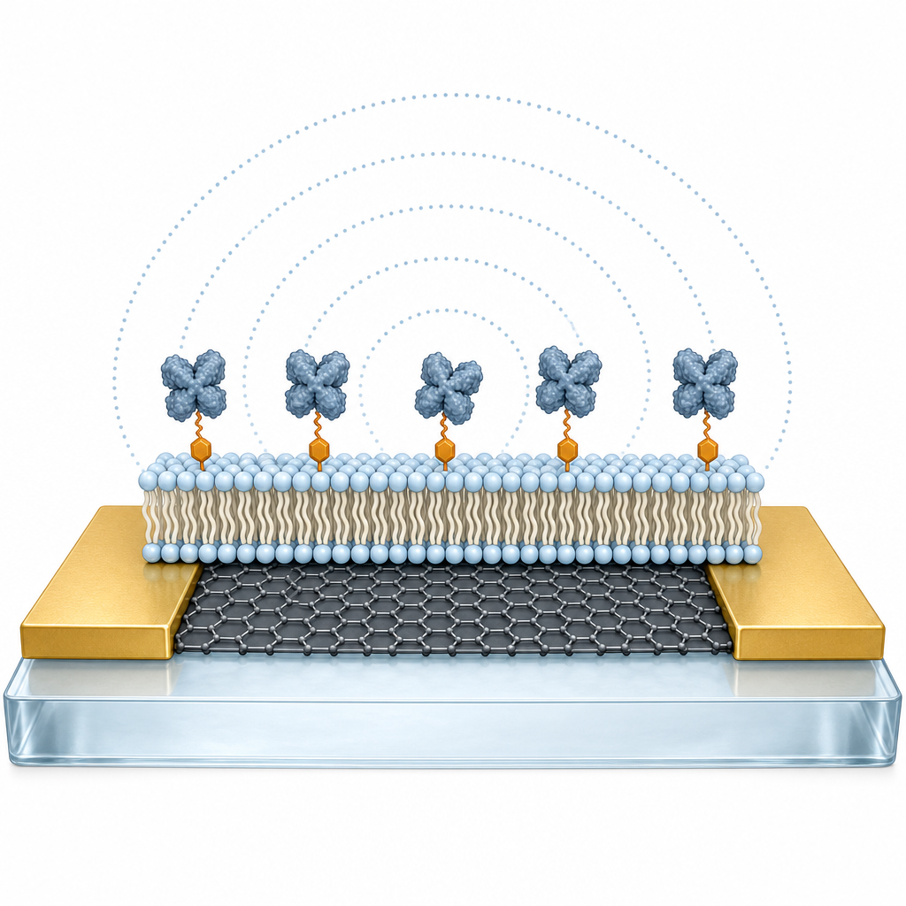
Using a modified fluorescence recovery after photobleaching (FRAP) method, which exploits ~5 µm laser-defined defects in the graphene where the otherwise-strong fluorescence quenching of graphene is locally suppressed, the authors directly confirmed that a continuous, laterally fluid SLB was present above the graphene/silica substrate. They found that fluid SLBs form readily by vesicle rupture when deposition occurs before an intermediate water layer has built up between graphene and the underlying silica; once this water layer is fully developed, the silica's affinity for rupturing vesicles is reduced and SLB formation is suppressed. To exploit both regimes, devices were vacuum-dried to remove the interfacial water before vesicle deposition; multiple gate-voltage sweeps were then applied to grow the water layer to a stable state before sensing. With this protocol, GFETs functionalized with 0–3 mol% biotin-lipids showed clear, dose-dependent shifts in the Dirac point upon exposure to 10 µg/mL Alexa-488–streptavidin in 1× PBS, while bovine serum albumin (BSA) controls produced negligible response, confirming receptor-specific detection. By varying ionic strength across 1×, 0.1×, 0.032× and 0.01× PBS the team probed Debye screening; comparison with a theoretical screening model showed that the SLB acyl-chain hydrophobic region contributes negligibly to ionic screening, so the effective detection region of the GFET extends beyond the bilayer and can capture binding events at the SLB's outer leaflet.
The combined membrane-coated GFET platform is directly relevant to label-free drug screening against G-protein-coupled receptors, ion channels and other membrane proteins, where preserving the lipid environment and receptor orientation is critical. It also enables studies of toxin–lipid interactions, raft formation and antibody–antigen recognition on cell-mimetic surfaces. Looking forward, the demonstration that FET sensitivity reaches above the bilayer suggests that engineered SLBs incorporating native or reconstituted membrane proteins could be coupled to GFET arrays for multiplexed electronic readouts, and that tuning ionic strength and lipid composition provides additional levers for controlling sensor response and selectivity in physiologically relevant buffers.
For researchers working on 2D biosensors, the central enabler here is the quality and transferability of the graphene channel. ACS Material's Trivial Transfer® Graphene allowed the team to obtain large-area, PMMA-supported CVD graphene that could be cleanly placed onto fused-silica device substrates and then liberated of its polymer carrier. Groups developing similar membrane-on-graphene biosensors, flexible bioelectronic interfaces or 2D-material FETs can source this same Trivial Transfer graphene, along with related CVD graphene-on-substrate options, from ACS Material's catalog to reproduce or extend this device architecture.How ACS Material products were used
- Trivial Transfer® Graphene (CVD graphene with PMMA support film) (Trivial Transfer Series) — “Trivial transfer CVD grapheneTM with poly(methyl methacrylate) (PMMA) films was purchased from ACS Material (Pasadena, CA).”
Product Performance in this StudyThe Trivial Transfer CVD graphene served as the active channel of the field-effect transistor. After PMMA removal and thermal annealing, it functioned as a high-mobility, biocompatible substrate onto which a fluid supported lipid bilayer was successfully assembled and used to detect streptavidin–biotin binding.
Related product categories
Frequently asked questionsCan a fluid supported lipid bilayer form on CVD graphene surfaces?
Yes. This study shows that a continuous, laterally fluid supported lipid bilayer (SLB) can form on CVD graphene by vesicle deposition, provided the intermediate water layer between graphene and the underlying silica has not yet developed. Vacuum-drying the graphene/silica device before adding vesicles preserves the silica's affinity for vesicle rupture, enabling reliable SLB formation that was verified by a defect-based FRAP method.
Why use Trivial Transfer CVD graphene for field-effect transistor biosensors?
Trivial Transfer CVD graphene from ACS Material is supplied with a PMMA support film that allows clean transfer onto custom-patterned silica or oxide substrates carrying pre-fabricated electrodes. This preserves the high carrier mobility of graphene, which is critical for sensitive FET biosensing, and avoids the carrier-mobility degradation and non-specific binding associated with direct chemical functionalization of the graphene channel.
How can a graphene FET detect binding events that occur beyond the Debye length?
The authors compared experimental Dirac-point shifts with a theoretical screening model across PBS dilutions from 1× to 0.01×. They found that the hydrophobic acyl-chain region of the supported lipid bilayer contributes negligibly to ionic screening. As a result, the effective detection region of the GFET extends beyond the 4–5 nm bilayer, enabling sensitive label-free detection of streptavidin binding to biotin-lipids at the outer leaflet.