-
Trivial Transfer Graphene for HER2 STEM Imaging - Leibniz INM, 2019
Jun 24, 2026 | ACS MATERIAL LLCPeckys, D. et al. (2019). Visualisation of HER2 homodimers in single cells from HER2 overexpressing primary formalin fixed paraffin embedded tumour tissue. *Molecular medicine*.
Molecular medicine · 2019
Researchers at Leibniz INM used ACS Material Trivial Transfer multilayer graphene to encapsulate tumour cells for STEM imaging of HER2 homodimers.
About this research
Researchers at INM – Leibniz Institute for New Materials used Trivial Transfer multilayer graphene from ACS Material LLC to encapsulate single tumour cells dissociated from formalin-fixed paraffin-embedded (FFPE) breast cancer biopsies, enabling scanning transmission electron microscopy (STEM) visualization of individually labelled HER2 receptors and statistical detection of HER2 homodimers in 33% and 55% of analysed images from two HER2 3+ patient samples. This work, published in Molecular Medicine (2019) by Peckys, Hirsch, Gaiser and de Jonge, demonstrates a route to extract functional receptor information - not just expression levels - directly from archived clinical biopsy material. The technique combines Affibody-biotin/streptavidin-quantum dot labelling with graphene-enclosed liquid-phase STEM.
HER2 (human epidermal growth factor receptor 2) is one of the most clinically important oncology biomarkers, used to stratify breast and gastric cancer patients for antibody therapies such as trastuzumab. Standard immunohistochemistry reports HER2 expression intensity but says nothing about the receptor's functional state. HER2 signalling, however, requires dimerization: the receptor spontaneously forms homodimers, and the homodimer fraction can differ markedly between cells and tumours. A method that reads out HER2 dimerization from routine FFPE specimens would close a meaningful gap between protein abundance and signalling competence, and could refine the selection of patients for targeted therapy. The technical hurdle has been imaging individual membrane proteins in hydrated cells at nanometre resolution while preserving compatibility with electron microscopy vacuum.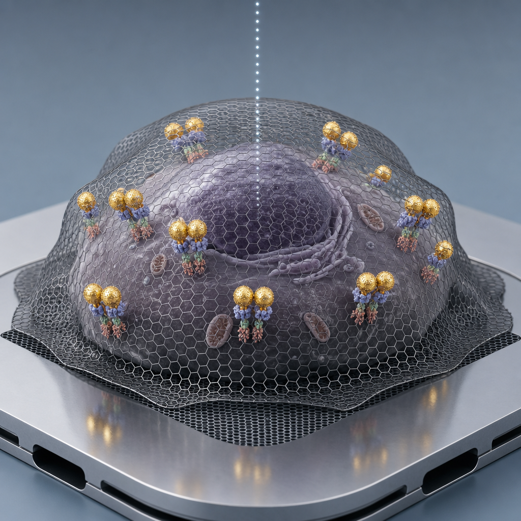
The ACS Material Trivial Transfer multilayer graphene (3-5 layers, supplied on a PMMA carrier) plays the central enabling role in this workflow. After labelling HER2 on dissociated tumour cells immobilized on silicon nitride microchips, the PMMA was stripped in acetone and the freestanding graphene sheets were floated on HPLC-grade water using a NaCl crystal carrier. A microchip held in curved tweezers was used to scoop up a floating sheet, which then sank onto the cells and conformed to their topography during air-drying. The graphene cover hermetically wraps each cell against the SiN membrane, keeping the cytoplasm and membrane hydrated even under the vacuum and 200 keV electron beam of an aberration-corrected STEM (JEOL ARM 200). Without this graphene encapsulation the cells would dehydrate and the QD label positions would shift, destroying single-molecule spatial information.
Key quantitative findings include membrane-bound HER2 surface densities ranging from 201 to 689 proteins/μm² across the analysed cells, consistent with HER2 3+ scoring. Dark-field STEM imaging at 60,000-120,000× magnification with pixel sizes between 0.66 and 1.66 nm and electron doses of 16-63 e⁻/Ų (below the radiation damage threshold for graphene-enclosed samples) yielded over 200,000 individually localized HER2-QD positions across the dataset. Automated ImageJ-based detection followed by pair correlation function g(r) analysis revealed HER2 homodimers in 33% of analysed images from patient 1 and 55% from patient 2, with no comparable peak in the HER2-negative control. The two-step Affibody-biotin / streptavidin-QD655 protocol gave clean 1:1 labelling stoichiometry. The total analysis time per patient image series was 20-30 minutes, suggesting reasonable throughput for translational use.
The approach is directly applicable to companion diagnostics for HER2-targeted therapies, where homodimer fraction may predict response to monoclonal antibodies and antibody-drug conjugates. More broadly, graphene-encapsulated liquid STEM extends to any membrane receptor that can be tagged with a quantum dot or comparable electron-dense label, including EGFR, MET, and other receptor tyrosine kinases relevant to oncology, as well as GPCR studies. The same graphene encapsulation strategy is finding use in cryo-correlative microscopy, in situ catalysis imaging, and protein-in-liquid TEM more generally. The authors point to extension toward heterodimer detection and toward routine biopsy analysis pipelines.
For researchers developing single-molecule electron microscopy workflows or hydrated-cell STEM, the Trivial Transfer multilayer graphene used here is available from ACS Material in formats designed for clean transfer to arbitrary substrates including SiN microchips. The product's combination of polymer carrier, water-floatable release, and conformal coverage of micron-scale biological objects is what makes the imaging protocol reproducible. The paper documents reliable cell-scale encapsulation at electron doses compatible with quantitative single-molecule localization, providing a useful reference point for groups planning similar in-liquid EM experiments on clinical specimens.How ACS Material products were used
- Trivial Transfer® Multilayer Graphene (Trivial Transfer Series) — “Trivial transfer multilayer graphene was purchased from, ACS Material LLC, Pasadena, CA, USA.”
Product Performance in this StudyThe multilayer graphene was used as a hydration-preserving cover over labelled tumour cells on SiN microchips, enabling liquid-phase STEM imaging of intact, hydrated cells. The graphene encapsulation worked reliably, allowing high-resolution detection of over 200,000 QD-labelled HER2 positions and successful pair-correlation analysis of HER2 homodimers.
Related product categories
Frequently asked questionsWhy is graphene used to encapsulate cells for STEM imaging?
Graphene encapsulation seals hydrated biological samples against the high vacuum and 200 keV electron beam of a scanning transmission electron microscope. A multilayer graphene sheet sinks onto cells on a silicon nitride membrane and conforms to their topography, preventing dehydration and stabilizing quantum-dot label positions. This allows single-molecule localization of membrane receptors such as HER2 with nanometre precision while avoiding the artefacts of conventional sample drying.
How does pair correlation function analysis detect HER2 homodimers?
After STEM imaging localizes individual quantum-dot-labelled HER2 receptors, their x,y coordinates feed into the pair correlation function g(r), which measures the probability of finding two labels at separation r relative to a random distribution. A peak at distances of roughly 10-20 nm above the random baseline indicates non-random clustering consistent with HER2 homodimers. The method handled over 200,000 label positions and found dimer signatures in 33-55% of images from HER2 3+ patients.
What grade of graphene is suitable for liquid-phase electron microscopy of cells?
Multilayer graphene with three to five layers on a PMMA carrier is generally preferred for cell encapsulation. The multilayer thickness gives mechanical robustness against tearing during the float-and-scoop transfer while remaining thin enough for high-contrast STEM imaging. The PMMA carrier enables clean water-surface transfer to silicon nitride microchips. The Trivial Transfer multilayer graphene used in this work fits these requirements and produced reproducible coverage of dissociated tumour cells.