-
Trivial Transfer Graphene for Liquid-Phase STEM - INM Leibniz, 2020
Jun 26, 2026 | ACS MATERIAL LLCWeinberg, F., Peckys, D. B., & Jonge, N. d. (2020). EGFR expression in HER2-driven breast cancer cells. *International Journal of Molecular Sciences*. https://doi.org/10.3390/ijms21239008
INM—Leibniz Institute for New Materials · International Journal of Molecular Sciences · 2020
Researchers at INM—Leibniz Institute for New Materials used ACS Material multi-layer trivial transfer graphene to image individual EGFR and HER2 receptors on breast cancer cells.
About this research
Researchers at INM—Leibniz Institute for New Materials, working with colleagues at Saarland University, used multi-layer trivial transfer graphene from ACS Material to seal hydrated SKBR3 breast cancer cells onto silicon nitride microchips for liquid-phase scanning transmission electron microscopy (STEM), enabling simultaneous single-molecule detection of EGFR and HER2 receptors in intact plasma membranes. The work, published in the International Journal of Molecular Sciences (2020), reports HER2 surface densities of 2–10 × 10² QDs/µm² and EGFR densities of 0.5–5 × 10¹ QDs/µm², and identifies a previously uncharacterized EGFR-enriched cell subpopulation comprising roughly 2.7% of the SKBR3 line. The graphene enclosure is central to the workflow: without it, hydrated cells cannot survive the electron beam and the high vacuum needed for nanometer-scale receptor mapping.
HER2 (ErbB2) is overexpressed in about 20% of breast cancers and is a major therapeutic target through agents such as trastuzumab, pertuzumab, lapatinib, and neratinib. Resistance to these therapies has been linked to heterodimerization of HER2 with other ErbB family members—particularly EGFR and HER3—but quantifying receptor stoichiometry at the single-cell, single-protein level remains difficult. Conventional approaches (FRET, proximity ligation, super-resolution microscopy, genetic tags) either lack spatial resolution, require exogenous overexpression, or produce false positives at the dense receptor populations typical of cancer cells. A label-and-image protocol that resolves individual receptors on native plasma membranes is therefore valuable for predicting therapy response and for identifying minority cell subtypes that may drive resistance.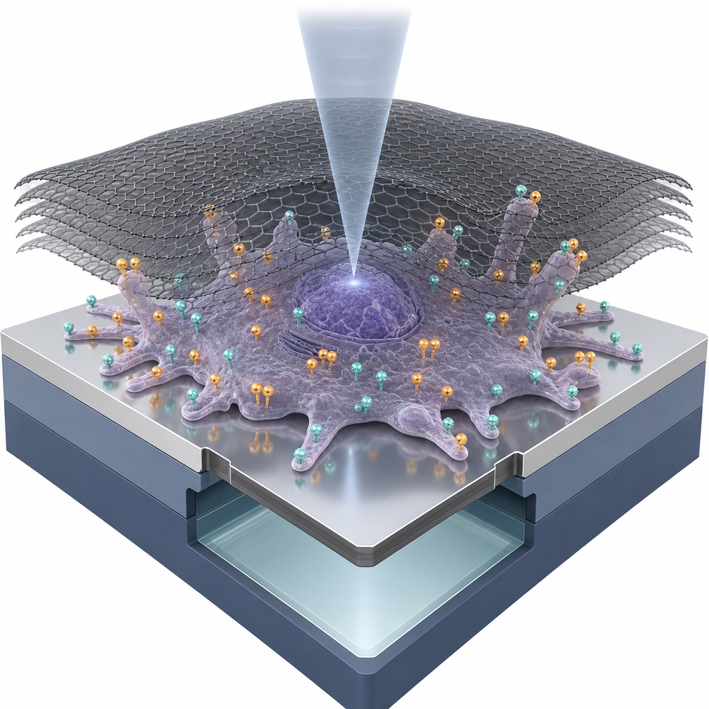
The ACS Material multi-layer graphene, supplied as polymethyl methacrylate-supported trivial transfer graphene ("Multi-layer graphene (polymethyl methacrylate trivial transfer graphene), (from ACS Materials, Pasadena, CA, USA)"), was floated off a NaCl substrate onto water, captured with a metal loop, and laminated over each microchip carrying SKBR3 cells that had been dual-labeled with biotinylated EGF/anti-HER2 Affibody and streptavidin-conjugated QD655/QD565. Excess water was wicked away with filter paper, producing a three- to five-layer graphene seal across the 50 nm SiN window. This thin, electron-transparent capping layer prevents dehydration and beam damage during STEM at 200 keV. The same chips could first be imaged by fluorescence microscopy (DIC and QD channels) and then transferred to the STEM, where overview brightfield images at M = 800× allowed re-identification of the optically scanned cells before high-resolution annular dark-field imaging at M = 120,000× (0.83 nm pixels, electron dose ≤280 e⁻Å⁻²).
The correlative workflow yielded quantitative receptor maps for every membrane region of interest. Across 31 bulk cells in clustered regions, HER2-QD565 averaged 2.7 ± 1.2 × 10² QDs/µm² and EGFR-QD655 averaged 0.6 ± 0.6 × 10¹ QDs/µm². Large membrane protrusions (LMPs) on bulk cells showed the highest HER2 density, 9.9 ± 4.5 × 10² QDs/µm², together with a roughly fourfold rise in EGFR to 2 ± 1 × 10¹ QDs/µm². Flat membrane regions carried the lowest density of both receptors (2.0 × 10² HER2 and 0.5 × 10¹ EGFR per µm²). A minority population (10 of 370 imaged cells, ~2.7%) was termed EGFR-enriched: on their LMPs, EGFR rose to 5 ± 2 × 10¹ QDs/µm², two- to threefold above bulk. HER2 exceeded EGFR by roughly a factor of 20 across the cell line, consistent with prior bulk transcriptomic data, while STEM revealed that EGFR is concentrated at signaling-active membrane ruffles rather than evenly distributed. Both single and dimeric EGFR particles were directly visible alongside HER2.
The ability to count individual receptors on unfixed-equivalent (chemically fixed but hydrated) cells has applications in HER2-targeted drug screening, identification of trastuzumab-resistant cancer stem-cell-like subpopulations, dimerization studies in patient-derived samples, and quantitative validation of FRET and proximity-ligation assays. The protocol could be extended to HER3 and HER4 with suitable affinity probes and, in principle, to any cell-surface receptor pair, enabling heterodimer mapping for bispecific antibody development. The authors specifically note that their dual-labeling and graphene-enclosure pipeline is compatible with primary tumor material, opening a route to single-cell molecular pathology of HER2-driven malignancies in bladder, prostate, and gastric cancers in addition to breast cancer.
For researchers building similar liquid-phase electron microscopy workflows, the multi-layer trivial transfer graphene used here is available from ACS Material as part of its Trivial Transfer Series. The PMMA support layer simplifies wet transfer onto small, fragile substrates such as SiN microchip windows, and the multi-layer format provides sufficient mechanical robustness to seal hydrated samples while remaining electron-transparent at 200 keV. The product is well suited to STEM, TEM, and graphene liquid cell experiments on biological specimens, electrocatalysts, and battery materials where preserving the native solvated state is essential.How ACS Material products were used
- Trivial Transfer® Graphene (multi-layer, PMMA-supported) (Trivial Transfer Series) — “Multi-layer graphene (polymethyl methacrylate trivial transfer graphene), (from ACS Materials, Pasadena, CA, USA).”
Product Performance in this StudyThe multi-layer trivial transfer graphene was used to enclose the silicon nitride microchips with adherent SKBR3 breast cancer cells, maintaining a hydrated sample state and protecting cells from electron-beam-induced damage during liquid-phase STEM imaging. This enclosure was essential to achieving single-protein resolution of QD-labeled EGFR and HER2 receptors.
Related product categories
Frequently asked questionsWhy is multi-layer graphene used to enclose cells for liquid-phase electron microscopy?
Multi-layer graphene seals hydrated cells onto a silicon nitride microchip, allowing them to remain in a liquid state inside the high-vacuum electron microscope and shielding them from electron-beam-induced radiolysis damage. Because graphene is only a few atoms thick, the electron beam still passes through with enough contrast to resolve single quantum-dot-labeled receptors at nanometer scale during scanning transmission electron microscopy.
How does trivial transfer graphene differ from CVD graphene on copper for biological imaging?
Trivial transfer graphene is supplied with a PMMA support layer that can be floated onto water and laminated directly over a fragile substrate such as a 50 nm SiN microscope window, without the etching, polymer coating, and rinsing steps required to release CVD graphene from copper foil. This makes it well suited to one-step enclosure of cells, electrochemical cells, and other hydrated samples for liquid-phase TEM and STEM.
What receptor densities did the study measure for HER2 and EGFR on SKBR3 cells?
HER2 ranged from 2 to 10 × 10² QDs/µm² across the SKBR3 plasma membrane, with the highest densities on large membrane protrusions of bulk cells (9.9 × 10² QDs/µm²). EGFR was approximately twentyfold lower, between 0.5 and 5 × 10¹ QDs/µm², and was concentrated on membrane protrusions and on a minority EGFR-enriched subpopulation comprising about 2.7% of imaged cells.