-
Trivial Transfer Graphene for Liquid-Phase STEM - Saarland University, 2021
Jun 30, 2026 | ACS MATERIAL LLCPeckys, D. B., Gaa, D., & Jonge, N. d. (2021). Quantification of EGFR-HER2 heterodimers in HER2-overexpressing breast cancer cells using liquid-phase electron microscopy. *Cells*. https://doi.org/10.3390/cells10113244
University Hospital · Cells · 2021
Saarland University used ACS Material Trivial Transfer Graphene to enclose breast cancer cells for liquid-phase STEM, quantifying EGFR-HER2 heterodimers.
About this research
Researchers at Saarland University used ACS Material multi-layer Trivial Transfer Graphene (3–5 layers, PMMA-supported) to enclose intact, chemically fixed HER2-overexpressing breast cancer cells for liquid-phase electron microscopy, enabling absolute single-molecule quantification of EGFR and HER2 receptors and their dimers. By combining correlative light microscopy (LM) and liquid-phase electron microscopy (LPEM) with two distinguishable quantum dots, the team detected more than 280,000 receptor positions across 41 SKBR3 cells. The work delivered absolute receptor surface densities, monomer/dimer percentages, and—most importantly—the subcellular heterogeneity of heterodimer shares across functionally distinct plasma-membrane regions, information that conventional pathology classification cannot capture.
This research matters because current breast cancer classification relies on coarse hormone-receptor and HER2 expression scoring, which misses the phenotypic complexity of tumors. EGFR and HER2 are overexpressed therapeutic targets across breast and gastric cancers, and their dimerization governs downstream tyrosine-kinase signaling that drives proliferation, survival, and drug resistance. Existing light-microscopy and immuno-gold approaches suffer from steric hindrance, label clustering, and excessive label-to-protein ratios at the high receptor densities (up to ~10^3/µm^2) found in overexpressing cells. A method that resolves individual receptors and directly measures physical dimerization in intact cells addresses a real gap in precision oncology, potentially identifying patients whose tumors carry an enhanced share of EGFR heterodimers and thus elevated risk of resistance to HER2-targeting antibody therapy.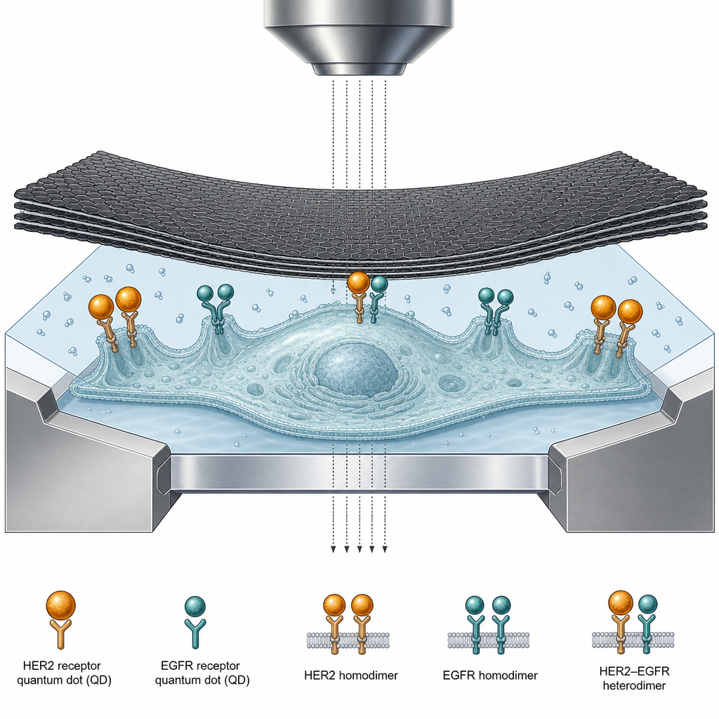
The ACS Material Trivial Transfer Graphene was central to sample preparation for the electron microscopy step. Immediately before imaging, microchips bearing dual-labeled cells were covered with the multi-layer graphene. The graphene sheet was released from its salt support and floated on water; a metal loop captured the floating film in a small droplet, and the microchip—cells facing the graphene—was positioned directly beneath, after which excess water was wicked away with filter paper. The 3–5 layer PMMA trivial transfer graphene thus formed a thin, electron-transparent, sealing enclosure over the hydrated cells on the 50 nm silicon-nitride window microchips. This enclosure preserved the liquid environment of the intact cells while permitting scanning transmission electron microscopy (STEM) at 200 keV beam energy on a JEOL ARM 200F. Imaging was performed at magnifications up to 120,000× (0.83 nm pixel size) with a calculated maximal electron dose of 280 e−/Ų, below the radiation-damage threshold for these samples, demonstrating that the graphene cover supported reliable, low-damage imaging of single quantum-dot labels.
Quantitatively, the labeling protocol used EGF-biotin with streptavidin-QD655 for EGFR and an anti-HER2 Affibody-biotin with smaller streptavidin-QD565 for HER2. Automated particle detection of STEM images yielded more than 9,300 labeled EGFR and over 275,000 labeled HER2 across 41 cells, with over 280,000 receptor positions analyzed overall. Pair correlation functions g(r) and bivariate g2(r) were used to extract homodimer and heterodimer fractions, validated against simulated images of known dimer percentages. Ligand-independent EGFR homodimers appeared at ~10% in all three membrane region types. HER2 homodimers rose from ~20% in lamellipodia to as high as 80% in large membrane protrusions (LMP). EGFR assembled in heterodimers at >40% in clustered regions and >80% in LMP, but only ~3% in lamellipodia; for HER2, the relative shortage of EGFR partners produced a maximum heterodimer share of ~6% in LMP. SKBR3 cells carry roughly 4-fold lower EGFR and ~50-fold higher HER2 expression than normal breast tissue. The analysis excluded distances below 7.5 nm to avoid counting overlapping nanoparticles, and statistical testing used one-way ANOVA with the Games–Howell post hoc test.
These findings enable a quantitative biomarker readout of receptor dimerization in intact single cells, distinct from relative methods such as the proximity ligation assay. The approach could help estimate total receptor numbers in patient tumors to optimize antibody-drug dosing, and could flag patients with elevated EGFR heterodimer shares who may need combination therapy—for example adding the EGFR inhibitor gefitinib or the irreversible ErbB inhibitor neratinib to standard trastuzumab and pertuzumab regimens. The authors note the method can be extended by swapping EGF or the anti-HER2 affibody for other binding ligands (e.g., IGF-I or an anti-HER3 affibody) to probe a broader range of membrane-protein interactions, and could be combined with their protocol for HER2 detection in dissociated FFPE tumor cells.
For researchers, this study illustrates the value of clean, transferable multi-layer graphene as a sealing window material for liquid-phase electron microscopy of biological specimens. The Trivial Transfer Graphene used here is available from ACS Material to groups developing correlative LM/LPEM workflows, in situ liquid-cell imaging, or graphene enclosure protocols for hydrated cells. Its ease of floating, capture, and transfer—performed under a simple binocular microscope—makes it well suited to single-molecule quantum-dot imaging where electron transparency and low radiation damage are essential.How ACS Material products were used
- Trivial Transfer® Graphene (multi-layer, 3–5 layers, PMMA) (Trivial Transfer Series) — “Multi-layer graphene (3–5 layers, polymethyl methacrylate trivial transfer graphene), from ACS Materials, Pasadena, CA, USA.”
Product Performance in this StudyThe PMMA-supported multi-layer Trivial Transfer Graphene was floated and laid over the silicon-nitride microchips to enclose intact, hydrated cancer cells, enabling liquid-phase STEM imaging at 200 keV without radiation damage at the applied dose. It performed as a reliable, sealing electron-transparent cover.
Related product categories
Frequently asked questionsWhy is multi-layer graphene used to enclose cells for liquid-phase electron microscopy?
Multi-layer graphene forms a thin, electron-transparent, sealing cover that preserves the liquid, hydrated state of intact cells while allowing scanning transmission electron microscopy. In this study, 3–5 layer Trivial Transfer Graphene was floated, captured with a metal loop, and laid over silicon-nitride microchips bearing labeled cells, enabling STEM imaging at 200 keV with a dose below the radiation-damage threshold.
How were EGFR-HER2 heterodimers quantified in breast cancer cells?
EGFR was labeled with EGF-biotin and streptavidin-QD655, and HER2 with an anti-HER2 Affibody-biotin and smaller streptavidin-QD565. Correlative light and liquid-phase electron microscopy located individual quantum dots, and pair correlation functions g(r) and bivariate g2(r) extracted homodimer and heterodimer fractions, validated against simulated images of known dimer percentages.
What were the key dimerization findings for EGFR and HER2 in SKBR3 cells?
Ligand-independent EGFR homodimers were about 10% across membrane regions. HER2 homodimers ranged from about 20% in lamellipodia to 80% in large membrane protrusions. EGFR heterodimer shares exceeded 40% in clustered regions and 80% in large membrane protrusions but only about 3% in lamellipodia, while HER2 heterodimers peaked near 6% due to the relative shortage of EGFR partners.