-
Graphene-Modified Paper Immunosensor for CRP — Chulalongkorn, 2019
Jun 24, 2026 | ACS MATERIAL LLCBoonkaew, S. et al. (2019). An origami paper-based electrochemical immunoassay for the C-reactive protein using a screen-printed carbon electrode modified with graphene and gold …. *Microchimica Acta*.
Microchimica Acta · 2019
Chulalongkorn researchers used ACS Material industrial graphene in an origami paper-based electrochemical immunoassay detecting C-reactive protein down to 15 ng/mL.
About this research
Researchers at Chulalongkorn University built an origami paper-based electrochemical immunoassay for C-reactive protein (CRP) using industrial-quality graphene powder purchased from ACS Material, LLC, combined with electrodeposited gold nanoparticles on a screen-printed carbon electrode (SPCE), and achieved a 15 ng mL⁻¹ limit of detection over the 0.05–100 μg mL⁻¹ range. The device folds together three paper panels carrying counter, reference, and graphene-modified working electrodes, integrating multi-step electrode modification, antibody immobilization, and impedimetric readout into a single disposable, low-cost format suitable for clinical screening of cardiovascular and inflammatory risk.
CRP is an acute-phase protein synthesized by the liver. Healthy serum levels stay below ~3 μg mL⁻¹, but during inflammation CRP can rise up to 1000-fold within 24–48 hours, signaling infection, sepsis risk, or elevated risk of cardiovascular events and heart attack. Conventional CRP assays — radial immunodiffusion, nephelometry, turbidimetry, ELISA, and radioimmunoassay — provide reliable quantification but rely on bulky instrumentation, large sample volumes, and trained operators. Point-of-care alternatives based on electrochemical paper devices address these constraints, but their sensitivity depends critically on the electrode material. Embedding high-surface-area, conductive carbon nanomaterials in the working electrode is one of the most direct ways to boost electron-transfer kinetics and lower detection limits without resorting to complex nanofabrication.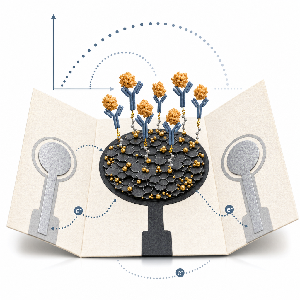
The ACS Material industrial graphene powder was used as the active filler in the working electrode ink. The authors prepared a graphene/carbon-ink composite at a 0.001:1 (w/w) graphene-to-carbon ratio, which was then screen-printed onto a wax-patterned Whatman No. 1 filter paper substrate to form the working electrode (5 mm diameter). Ag/AgCl ink was screened separately for the reference electrode and conductive pads. After baking at 55 °C for 1 hour, gold nanoparticles were electrodeposited onto the graphene-modified SPCE from 5 mM K[AuCl₄] in 0.5 M H₂SO₄ at −0.5 V for 300 s. A self-assembled monolayer of L-cysteine was then formed overnight at 4 °C, activated with EDC/NHS, and covalently coupled to anti-CRP capture antibody (50 μg mL⁻¹). BSA blocked remaining sites. The graphene layer thus served two roles: it increased the electroactive surface area available for AuNP nucleation and enhanced charge-transfer efficiency between the electrode and the [Fe(CN)₆]³⁻/⁴⁻ redox probe used in EIS.
Electrochemical characterization confirmed that graphene incorporation lowered the charge-transfer resistance compared with bare SPCE, and SEM/EDS verified successful stepwise deposition of AuNPs, L-cysteine, and antibody. Under optimized conditions — incubation at 4 °C for 50 min, EIS measurement at 0.1 V with 0.01 V amplitude over 100 kHz to 0.01 Hz in 5 mM [Fe(CN)₆]³⁻/⁴⁻/0.1 M KNO₃ — the charge-transfer resistance increased linearly with CRP concentration across 0.05 to 100 μg mL⁻¹, spanning the clinically relevant range from healthy baseline through acute inflammatory states. The limit of detection was 15 ng mL⁻¹ at S/N = 3, well below the ~3 μg mL⁻¹ healthy cutoff. The platform was validated using a certified reference human serum (ERM-DA474/IFCC, 41.2 μg mL⁻¹) diluted to 5–40 μg mL⁻¹, with recoveries in good agreement with the certified value. Triplicate measurements gave reproducible responses, and the device was stable when stored at 4 °C between modification and use.
The origami architecture enables sequential modification on one folded face and detection on another, avoiding cross-contamination of the working electrode during antibody immobilization. This design choice, combined with the low material cost of paper, screen-printable carbon ink, and bulk graphene powder, points toward viable disposable point-of-care diagnostics for CRP screening in resource-limited settings, primary care offices, and home testing. The same architecture can be repurposed for other antibody-based assays — cardiac troponins, cytokines, infectious disease biomarkers — and the graphene/AuNP working electrode is compatible with aptamer or molecularly imprinted polymer recognition layers, extending its scope to small-molecule biosensing and environmental monitoring.
For researchers developing paper-based or screen-printed biosensors, the takeaway is that bulk, industrial-grade graphene powder provides a cost-effective performance boost when mixed into carbon inks, without requiring single-layer flakes or specialty pastes. ACS Material supplies industrial-quality graphene and graphene nanoplatelet powders suitable for ink formulation, electrode coating, and composite fabrication in immunoassay, electrochemical sensing, and energy storage research.How ACS Material products were used
- Industrial Thin Layer Graphene Nanoplatelets (Graphene Series) — “Industrial-quality graphene powder was purchased from ACS material, LLC (Pasedena, USA, https://www.acsmaterial.com).”
Product Performance in this StudyThe industrial-grade graphene was blended with carbon ink (0.001:1 w/w) to fabricate the graphene-modified screen-printed carbon electrode (G/SPCE), enhancing electron transfer and sensitivity for impedimetric CRP detection. It enabled a wide linear range (0.05–100 μg mL⁻¹) and a low limit of detection of 15 ng mL⁻¹.
Related product categories
Frequently asked questionsWhy use graphene in a screen-printed carbon electrode for biosensing?
Adding graphene to carbon ink increases the electroactive surface area and lowers charge-transfer resistance between the electrode and the redox probe. In this CRP immunosensor, the graphene-modified SPCE enabled detection down to 15 ng mL⁻¹ — well below the ~3 μg mL⁻¹ healthy serum threshold — and provided a linear impedance response across the clinically relevant 0.05–100 μg mL⁻¹ range using [Fe(CN)₆]³⁻/⁴⁻ as the redox couple.
How is industrial graphene powder mixed into screen-printable carbon ink?
The authors blended ACS Material industrial-quality graphene powder with carbon ink at a 0.001:1 (w/w) ratio. The composite was screen-printed onto wax-patterned Whatman No. 1 filter paper to form a 5 mm working electrode, then baked at 55 °C for 1 hour to remove solvent. This simple bulk-dispersion approach avoids specialty graphene pastes while still delivering measurable sensitivity gains over unmodified carbon SPCEs.
What is the clinical detection range and limit for paper-based CRP immunoassays?
This origami paper device covers 0.05 to 100 μg mL⁻¹ of C-reactive protein with a 15 ng mL⁻¹ detection limit at S/N = 3. The range spans healthy baselines (<3 μg mL⁻¹) and elevated inflammatory levels associated with cardiovascular and infection risk. The method was validated against a certified reference human serum (ERM-DA474/IFCC, 41.2 μg mL⁻¹) diluted into the 5–40 μg mL⁻¹ working window.