-
Graphene Nanoplatelet Laccase Biosensor - Karl-Franzens Univ. Graz, 2020
Jun 23, 2026 | ACS MATERIAL LLCZrinski, I. et al. (2020). Evaluation of phenolic antioxidant capacity in beverages based on laccase immobilized on screen-printed carbon electrode modified with graphene nanoplatelets and gold nanoparticles. *Microchemical Journal*. https://doi.org/10.1016/j.microc.2019.104282
Microchemical Journal · 2020
Karl-Franzens University of Graz researchers used ACS Material graphene nanoplatelets (2–10 nm) to build a laccase biosensor detecting phenolic antioxidants in wine.
About this research
Researchers at Karl-Franzens University of Graz, working with collaborators in Zagreb, Bangkok, Ljubljana and Belgrade, used graphene nanoplatelets (2–10 nm thickness) supplied by ACS Material to construct a laccase-based electrochemical biosensor (LACC/AuNP/GNPl/SPCE) that quantifies phenolic antioxidant capacity in wine and blueberry syrup. Published in Microchemical Journal (2020), the work demonstrates that integrating these graphene nanoplatelets with gold nanoparticles in a screen-printed carbon electrode produces a hydroquinone biosensor with a 1.5 µM detection limit, a 4–130 µM linear range and antioxidant capacity values that match the reference Trolox Equivalent Antioxidant Capacity (TEAC) spectrophotometric assay.
Phenolic antioxidants in foods and beverages are central to discussions of cardiovascular health, anticancer activity and oxidative stress mitigation. Established quantification routes—Folin–Ciocalteu spectrophotometry, HPLC and capillary electrophoresis coupled to UV, fluorescence or mass spectrometry—are accurate but slow, expensive and demand sample pre-concentration. Electrochemical biosensors built on screen-printed electrodes offer a low-cost, rapid alternative, yet their sensitivity depends critically on the electrode matrix. Bare carbon surfaces suffer from signal fouling, enzyme denaturation and poor reproducibility. Adding nanomaterial modifiers can shorten electron transfer distances to enzyme redox centers and preserve enzyme orientation. The authors therefore set out to engineer a biosensor matrix where graphene nanoplatelets and gold nanoparticles work synergistically to stabilize immobilized laccase and amplify the current generated when phenolic substrates are catalytically oxidized.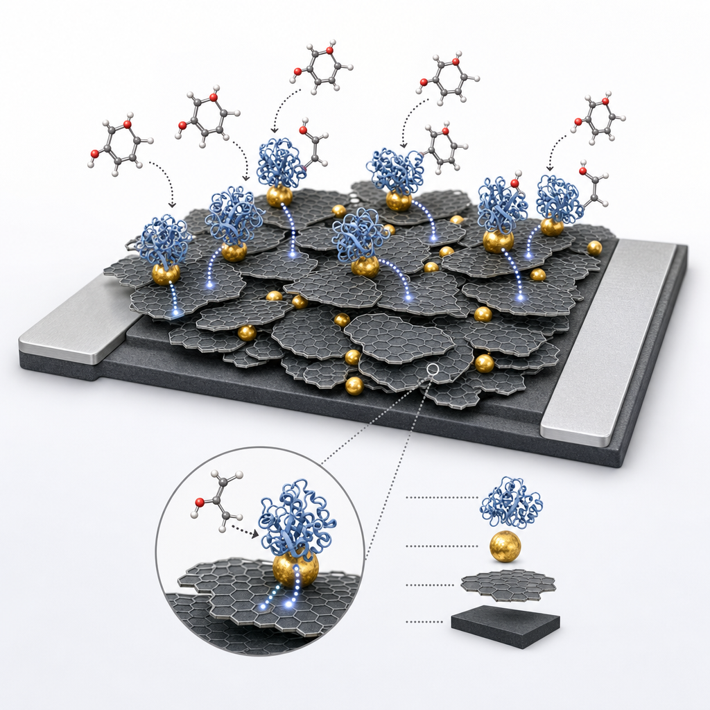
The ACS Material graphene nanoplatelets entered the workflow at the electrode fabrication step. The Methods section specifies the modifier as "graphene nanoplatelets (5% m:m; 2–10 nm thickness; GNPlt 2-10–50 g; ACS Material, USA)," mixed directly into a Gwent carbon ink (C2030519P4). The graphene-loaded ink was screen-printed onto pre-etched alumina substrates using a semi-automatic MPM SP-200 printer and dried at room temperature. A silver conductive paint provided the contact pad, and the active area was defined at 0.28 cm² with nail enamel insulation. The GNPl loading was optimized between 1% and 10% m:m by cyclic voltammetry; 5% gave the strongest cathodic peak current without the agglomeration losses observed at 10%. Onto this GNPl/SPCE substrate the authors drop-cast 5 µL of 5 nm gold nanoparticle suspension, followed by 5 µL of a laccase cocktail containing Nafion neutralized with ammonia, enzyme solution (1 mg/0.5 mL phosphate buffer), ethanol and water. SEM imaging confirmed that GNPl produced a flaky, homogeneous surface covering insulating zones of bare SPE, while gold nanoparticles ensured uniform enzyme distribution.
The optimized LACC/AuNP/GNPl/SPCE detects hydroquinone by hydrodynamic amperometry at –0.05 V vs Ag/AgCl in 0.1 M phosphate buffer (pH 7.0). Calibration was linear from 4 to 130 µM with R² = 0.9987, a 1.5 µM detection limit (3σ) and a 5 µM quantification limit. Repeatability over five measurements at 100 µM hydroquinone was ±2% RSD, and reproducibility across five independently fabricated biosensors was ±3% RSD—a clear improvement on the ~10% RSD observed without GNPl. Cyclic voltammetry showed Ipa and Ipc scaling linearly with √ν (R² = 0.9887 and 0.9944), indicating diffusion-controlled kinetics. Interference screening with ascorbic acid, glucose, dopamine and paracetamol gave responses below 10% of the hydroquinone signal, while phenolic analytes such as caffeic acid, p-coumaric acid, sinapic, syringic and ferulic acids yielded >23% relative responses, confirming broad polyphenol selectivity. Applied to commercial wine and blueberry syrup, the biosensor returned Trolox-equivalent values of 36.9 ± 0.2 µM (wine) and 77.6 ± 0.3 µM (syrup), in close agreement with the TEAC spectrophotometric assay (37.2 ± 0.2 and 79.9 ± 0.6 µM respectively). Stability remained intact after five days of use.
The resulting platform is directly relevant to quality control in the wine, fruit liquor, juice and functional-beverage industries, where phenolic antioxidant capacity is a routine release specification. The same chemistry extends to pharmaceutical formulations containing polyphenolic excipients, cosmetic antioxidant claims and environmental monitoring of phenolic pollutants in wastewater. The authors explicitly point to extending the biosensor to human blood samples as a future direction, opening a path toward clinical antioxidant status assays. Because the device is screen-printed and disposable, it is amenable to portable, point-of-need field testing—useful for vineyard producers, beverage QA labs and regulatory inspectors who currently rely on benchtop UV-Vis instrumentation and the time-consuming Folin–Ciocalteu protocol.
For researchers developing similar enzymatic or electrocatalytic sensors, the graphene nanoplatelets used here are available through ACS Material's Graphene Series with thickness grades including 2–10 nm. The paper's data show that selecting the right thickness and loading is decisive: 5% m:m delivered optimal current enhancement, while higher loadings caused agglomeration-driven losses. Combined with gold nanoparticles, the GNPl matrix preserved laccase activity at physiological pH and produced a calibration window broad enough to bracket real beverage samples without dilution. Groups working on phenol biosensors, food-analysis electrochemistry or laccase-based bioelectrocatalysis will find these graphene nanoplatelets a practical building block, supported by the quantitative performance documented in this study.How ACS Material products were used
- Graphene Nanoplatelets (2-10nm) (Graphene Series) — “modified with graphene nanoplatelets (5% m:m; 2–10 nm thickness; GNPlt 2- 10–50 g; ACS Material, USA)”
Product Performance in this StudyGraphene nanoplatelets (2–10 nm) from ACS Material were blended at 5% (m:m) into the carbon ink to fabricate the screen-printed electrode. Their high conductivity and large surface area sharpened the hydroquinone redox peaks and improved repeatability from ~10% RSD to ~2% RSD, anchoring the analytical performance of the laccase biosensor.
Related product categories
Frequently asked questionsHow do graphene nanoplatelets improve laccase biosensor performance?
Graphene nanoplatelets act as conductive electronic wires that shorten the electron transfer distance between the laccase active site and the electrode while protecting the enzyme from adsorptive denaturation. In this study, mixing 5% m:m of 2–10 nm ACS Material graphene nanoplatelets into the carbon ink raised the hydroquinone cathodic peak current and improved measurement repeatability from about 10% RSD to roughly 2% RSD across five replicates.
What detection limit was achieved for hydroquinone with this biosensor?
The LACC/AuNP/GNPl/SPCE biosensor reached a detection limit of 1.5 µM (3σ) for hydroquinone, with a quantification limit of 5 µM and a linear amperometric response from 4 to 130 µM in 0.1 M phosphate buffer (pH 7.0) at an operating potential of –0.05 V versus Ag/AgCl. Repeatability was ±2% RSD and reproducibility across five biosensors was ±3% RSD.
Why combine gold nanoparticles with graphene nanoplatelets in a screen-printed electrode?
Graphene nanoplatelets provide high conductivity and a large active surface, while gold nanoparticles add metallic conductivity, lower charge transfer resistance, and offer preferential adsorption sites that distribute laccase homogeneously across the electrode. Together they synergistically increase the redox current for hydroquinone, enabling stable, reproducible amperometric detection at –0.05 V with negligible interference from ascorbic acid, glucose, dopamine or paracetamol.