-
Graphene Oxide ATP Biosensor for Plants - University of Florida, 2015
Jun 08, 2026 | ACS MATERIAL LLCVanegas, D. C. et al. (2015). A self-referencing biosensor for real-time monitoring of physiological ATP transport in plant systems. *Biosensors and Bioelectronics*. https://doi.org/10.1016/j.bios.2015.05.027
Biosensors and Bioelectronics · 2015
University of Florida researchers built a self-referencing ATP micro-biosensor on an rGO/nanoplatinum electrode using ACS Material single-layer graphene oxide, reaching a 1.3 nM detection limit.
About this research
Researchers at the University of Florida, together with collaborators at the University of Texas at Austin and the University of Colorado Denver, used single-layer graphene oxide from ACS Material to fabricate a self-referencing electrochemical micro-biosensor that directly measures extracellular ATP (eATP) flux near living plant cells, achieving a sensitivity of 2.4 ± 1.8 nA/μM and a lower detection limit of 1.3 ± 0.7 nM. The device combines a reduced graphene oxide / nanoplatinum (rGO/nPt) transducer with a stratified bi-enzyme nanocomposite of glycerol kinase (GK) and glycerol-3-phosphate oxidase (G3PO) immobilized in a laponite hydrogel and protein-templated silica sol–gel. Published in Biosensors and Bioelectronics in 2015, the work demonstrates the first non-invasive ATP micro-biosensor capable of resolving the temporally transient nanomolar movement of ATP at physiologically relevant levels.
Extracellular ATP is a hormone-like signaling molecule that regulates diverse responses in plant and animal cells, including growth, wounding response, and gravity-directed polarization. Existing tools — primarily luciferin–luciferase bulk assays and earlier amperometric devices based on hexokinase/glucose oxidase — either consume the sample, suffer from glucose interference, or lack the spatial and temporal resolution needed to follow ATP transport in real time near the cell surface. Capturing nM-level ATP gradients in the unstirred layer adjacent to plant tissues is critical for understanding purinergic signaling, apyrase-mediated hydrolysis, and stress responses such as wounding and touch. A reversible, flux-sensitive probe that can be operated in self-referencing mode addresses this gap and complements optical assays with quantitative directional information.
The ACS Material single-layer graphene oxide (thickness 0.8–1.2 nm, 99% purity, modified Hummers method) is the structural foundation of the transducing layer. A 2 mg/mL aqueous GO solution was ultrasonicated for 30 minutes, then partially reduced by adding ascorbic acid at 8 mg/mL. The Pt/Ir microelectrode tip (1–2 µm diameter) was dip-coated in this rGO suspension and dried, after which nanoplatinum clusters were sonoelectrodeposited on top from a chloroplatinic acid / lead acetate bath at 10 V for 30 s. A Nafion membrane was applied for charge and size exclusion of interferents. The rGO/nPt surface was then sequentially loaded with laponite hydrogels containing G3PO and GK, with each enzyme layer encapsulated by a protein-templated silica nanoshell (~100 nm thick, ~10 nm pores) formed from TEOS sol–gel. The high electroactive area of the rGO/nPt platform, derived from the ACS Material GO, enabled larger enzymatic loading than carbon-nanotube-based platforms reported by the same group.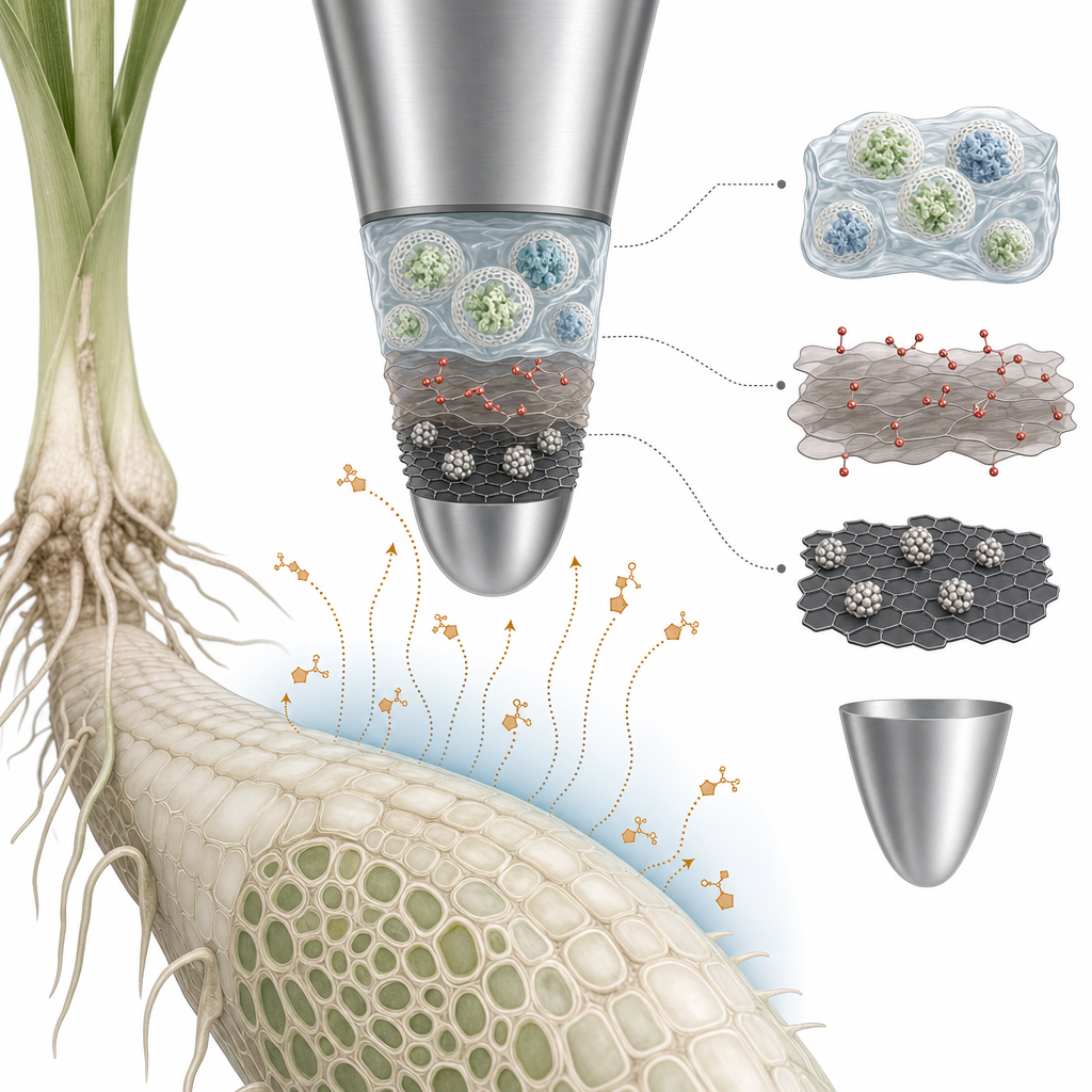
Electrochemical characterization by DC potential amperometry at +700 mV vs. Ag/AgCl gave a sensitivity of 2.4 ± 1.8 nA/μM, a 95% response time of 20 ± 13 s, a lower detection limit of 1.3 ± 0.7 nM, and a signal-to-noise ratio ≥3 over an operating range of 1.3 nM to 12 μM. Sensitivity decreased by only 1.33 ± 0.52% over seven days. Abiotic validation against a 15 mM ATP point source in 2% agarose showed a correlation coefficient of 0.98 between measured flux and a Fickian diffusion model. In Zea mays roots, baseline eATP was 2.6 ± 0.3 nM; wounding raised it to 3.4 ± 0.1 μM, and touch stimulation produced a ~4-fold rise to 10.4 ± 2.9 nM. In germinating Ceratopteris richardii spores, the sensor resolved oscillatory ATP release reaching 5.0–6.5 nM in 10–12 minute cycles, with a net efflux of 13.2 ± 4.8 pmol-ATP h⁻¹.
This platform opens quantitative, real-time investigation of purinergic signaling in plant physiology — root development, gravitropism, wounding, and touch responses — and is applicable to any system where nanomolar eATP plays a role, including mammalian cells, bacterial biofilms, and tissue culture. Comparable luciferase data place eATP at 2–4 nM in healthy Arabidopsis root media, 28 nM–1 μM in human plasma, and up to 32 nM in oral Candida albicans biofilms, all within this sensor's operating window. Self-referencing flux measurement adds directional information that bulk assays cannot provide, enabling researchers to map dynamic ATP transport at the cell surface. The authors point to future use in studies of apyrase activity, purinoceptor agonist/antagonist screening, and stress-induced eATP release in crop species.
For researchers building enzyme-based electrochemical biosensors, the role of single-layer graphene oxide here is instructive: a thin, high-purity GO flake reduced in situ produces a conductive scaffold with enough surface area to host a stratified bi-enzyme assembly and still resolve sub-nanomolar analytes. The graphene oxide and related graphene products used in this study are available from ACS Material under the Graphene Series, supporting groups working on amperometric biosensors, flexible bioelectrodes, neurochemical probes, and nanocomposite electrochemical platforms for life-science research.How ACS Material products were used
- Single Layer Graphene Oxide Flake (H Method) (Graphene Series) — “Single-layer graphene oxide (GO) (Thickness: 0.8 to 1.2 nm; purity: 99%; manufacturing method: modified Hummer) was procured from ACS Material (Medford, MA).”
Product Performance in this StudyThe ACS Material single-layer graphene oxide was partially reduced with ascorbic acid to form an rGO/nanoplatinum platform on the microelectrode tip. This nanocomposite provided high electroactive surface area and enabled larger enzyme loading, supporting the biosensor's nanomolar detection limit and stable response over a seven-day study.
Related product categories
Frequently asked questionsWhy use single-layer graphene oxide in an ATP biosensor electrode?
Single-layer graphene oxide reduced with ascorbic acid produces a thin, highly conductive rGO scaffold with abundant surface area and oxygen functionalities. Combined with sonoelectrodeposited nanoplatinum, this rGO/nPt platform offers a large electroactive area that supports higher enzyme loading than carbon nanotube platforms, enabling sub-nanomolar ATP detection (1.3 nM limit) while keeping response time near 20 seconds.
How does a self-referencing biosensor measure ATP flux rather than concentration?
The microelectrode oscillates over a fixed excursion distance perpendicular to the cell surface, sampling current at near and far poles. The differential current is converted to a concentration gradient and applied to Fick's first law of diffusion using ATP's diffusion constant (2.6 × 10⁻⁶ cm² s⁻¹). This yields directional flux data and discriminates true cellular transport from background drift.
What detection limit is achievable for extracellular ATP in plant tissue?
The stratified bi-enzyme micro-biosensor built on an rGO/nanoplatinum platform achieved a 1.3 ± 0.7 nM lower detection limit with a 20 ± 13 s response time and a linear range up to 12 μM. This is sufficient to resolve baseline eATP near Zea mays roots (~2.6 nM) and oscillatory release from germinating Ceratopteris spores (1.5–6.5 nM cycles).