-
Graphene Oxide Fiber Microelectrodes for Neurochemical Sensing - University of Cincinnati, 2022
Jun 18, 2026 | ACS MATERIAL LLCLi, Y. et al. (2022). Graphene-fiber microelectrodes for ultrasensitive neurochemical detection. *Analytical Chemistry*. https://doi.org/10.1021/acs.analchem.1c05637
University of Cincinnati · Analytical Chemistry · 2022
University of Cincinnati researchers used ACS Material single-layer graphene oxide dispersion to spin GO/rGO fiber microelectrodes that detect dopamine down to 61 nM.
About this research
Researchers at the University of Cincinnati used ACS Material single-layer graphene oxide (GO) dispersion to fabricate graphene-fiber microelectrodes (GFMEs) that achieved subsecond neurochemical detection with fast-scan cyclic voltammetry (FSCV) for the first time, including dopamine detection limits near 61 nM. The team produced both GO and reduced graphene oxide (rGO) microfibers, roughly 20–30 µm in diameter, through a one-step dimension-confined hydrothermal strategy. Compared with traditional carbon-fiber microelectrodes (CFMEs), these graphene fibers showed faster electron transfer kinetics, enhanced redox cycling, higher sensitivity, frequency-independent behavior, and improved resistance to chemical fouling, demonstrating their value as biological sensors.
This research addresses a long-standing limitation in neurochemical monitoring. FSCV at carbon-fiber electrodes is the dominant approach for measuring electroactive neurotransmitters such as dopamine and serotonin in real time, but the heterogeneous, hard-to-tune surface of carbon fibers limits how precisely electrode chemistry and structure can be controlled. Newer carbon nanomaterials such as carbon nanotube fibers improve performance but are expensive and require specialized reactors or synthesis expertise, putting them out of reach for many laboratories. There is therefore a clear need for electrode materials that combine excellent electrochemical properties with simple, accessible fabrication and homogeneous, tunable surfaces. Pure graphene fibers, made directly from graphene oxide, allow direct study of electrode–analyte interactions without interference from an underlying substrate, which matters for biosensing, neuroscience, and electroanalytical chemistry research.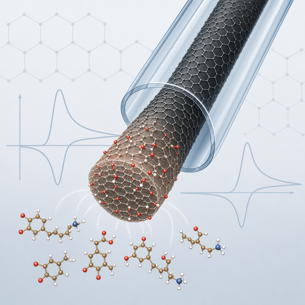
The ACS Material product served as the essential starting material. The Methods section states that a single-layer graphene oxide dispersion in water at 10 mg/mL was obtained from ACS Material (Pasadena, CA). To form fibers, the team mixed the 10 mg/mL GO dispersion with 1 w/w% L-ascorbic acid, injected the suspension into a thin glass capillary, sealed the ends with epoxy, and heated it in an oven. Treatment at 80 °C for 2 hours produced GO microfibers, while heating at 280 °C for 1 hour reduced the GO to rGO fibers. The ascorbic acid acted as a binder to strengthen π–π interactions between GO sheets, enabling robust fiber assembly at low temperature. Fibers were released in isopropyl alcohol, aspirated into a second capillary, pulled, sealed, and polished into disk microelectrodes. The graphene fibers were then characterized by SEM, Raman spectroscopy, and XPS, and tested electrochemically with both slow-scan cyclic voltammetry and FSCV against redox probes and real neurochemicals.
Quantitatively, XPS showed oxygen content dropping from 30.1% on GO fibers to 15.1% on rGO fibers after thermal reduction, a roughly two-fold reduction confirming distinct surface chemistries. Raman analysis gave ID/IG ratios of 1.98 for GO and 1.67 for rGO, and ID″/IG ratios of 0.30 and 0.40 respectively, indicating different defect levels and crystallinity. Dopamine sensitivity reached 1.54 ± 0.5 nA/µM at GO and 1.35 ± 0.4 nA/µM at rGO microelectrodes, far above the 0.41 ± 0.09 nA/µM measured at bare disk carbon-fiber electrodes. Limits of detection for dopamine were 61.1 ± 1.4 nM (GO), 68.6 ± 2.3 nM (rGO), and 185.2 ± 10.4 nM (carbon fiber). Both graphene fibers exhibited smaller peak separations (ΔEp) and reduction-to-oxidation current ratios near 0.77 (GO) versus 0.39 for carbon fiber, indicating faster, more reversible electron transfer. The fibers maintained frequency-independent behavior, losing only 14.1% (GO) and 33.1% (rGO) of oxidative current at 100 Hz versus 68.3% for carbon fiber, supporting temporal resolution down to 10 ms. For serotonin antifouling, GO and rGO retained roughly 103–104% of signal after 25 injections while carbon fiber lost 52.3%. Endogenous dopamine release was successfully measured ex vivo in rat caudate-putamen slices.
These results enable more accessible, tunable electrodes for real-time neurochemical sensing. Because the fibers can be synthesized in a standard laboratory without expensive reactors, they lower the barrier for researchers in neuroscience, electroanalytical chemistry, and biosensing who want to study dopamine, serotonin, norepinephrine, adenosine, and guanosine. The ability to switch between GO and rGO chemistries allows surfaces to be matched to specific analytes, and disk-format microelectrodes promise improved spatial resolution for tissue measurements compared with conventional cylindrical carbon fibers. The authors point to future work on controlling the 3D porosity of the fibers to further tune catecholamine cyclization and frequency behavior, and on extending these electrodes to additional analytes and in vivo applications.
For researchers working on similar electrochemical sensors, the single-layer graphene oxide dispersion used here is available from ACS Material in the Graphene Series. The paper demonstrates that a well-characterized, water-based GO dispersion can serve directly as the feedstock for fiber-spun microelectrodes with reproducible, high-quality electrochemical performance. This makes such GO dispersions a practical building block for neurochemical microsensors, flexible bioelectronics, and other applications where tunable, low-defect graphene surfaces are needed.How ACS Material products were used
- Single Layer Graphene Oxide Dispersion (10 mg/mL in water) (Graphene Series) — “Single-layer graphene oxide dispersion in water (10 mg,mL, ACS material, Pasadena, CA)”
Product Performance in this StudyThe ACS Material single-layer GO dispersion was the sole feedstock for hydrothermally spun GO and rGO microfibers, which delivered higher dopamine sensitivity, lower detection limits, and superior antifouling versus carbon-fiber electrodes.
Related product categories
Frequently asked questionsHow does graphene oxide improve dopamine detection compared to carbon fiber?
In this study, graphene oxide fiber microelectrodes showed dopamine sensitivity of 1.54 nA/µM versus 0.41 nA/µM for bare carbon fiber, and a detection limit of 61 nM versus 185 nM. The graphene oxide surface gave faster electron transfer, more reversible redox cycling, and oxide functionality that promotes dopamine adsorption, all of which raise sensitivity and lower detection limits.
What is single-layer graphene oxide dispersion used for in microelectrode fabrication?
The single-layer graphene oxide dispersion served as the direct feedstock for spinning fibers. Researchers mixed the 10 mg/mL aqueous dispersion with ascorbic acid, injected it into a glass capillary, and heated it hydrothermally. Heating at 80 °C produced graphene oxide fibers, while 280 °C reduced them to rGO fibers, which were then polished into disk microelectrodes for neurochemical sensing.
Why is frequency-independent behavior important for neurochemical sensors?
Frequency-independent behavior lets electrodes maintain sensitivity at higher scan frequencies, improving temporal resolution for tracking fast neurotransmitter changes. In this work, graphene oxide fibers lost only 14.1% of dopamine oxidative current at 100 Hz, compared with 68.3% for carbon fiber, indicating that measurements with temporal resolution down to 10 ms are achievable.