-
Graphene Oxide FRET Biosensor for Antibiotic Resistance - WKU, 2023
Jun 18, 2026 | ACS MATERIAL LLCKang, J., Nguyen, V., & Kim, M. (2023). Rapid and sensitive detection of antibiotic resistance genes by utilizing TALEs as a diagnostic probe with 2D-nanosheet graphene oxide. *Analytical Chemistry*. https://doi.org/10.1021/acs.analchem.3c00647
Western Kentucky University · Analytical Chemistry · 2023
Western Kentucky University used ACS Material graphene oxide as a FRET quencher with QD-labeled TALEs to detect tetM antibiotic resistance genes at 1 fM in 10 minutes.
About this research
Researchers at Western Kentucky University developed a rapid, amplification-free biosensor that combines graphene oxide (GO) dispersion from ACS Material with quantum-dot-labeled transcriptional activator-like effectors (TALEs) to detect the tetracycline resistance gene tetM at a limit of detection as low as 1 fM of Staphylococcus aureus genomic DNA. The system exploits GO as an efficient fluorescence quencher: QD-labeled TALEs adsorb onto the GO nanosheet surface, quenching the quantum dot emission by fluorescence resonance energy transfer (FRET). When the TALE binds its target double-stranded DNA, it dissociates from GO, restoring the signal in a turn-on fashion. The whole assay runs at room temperature in a one-pot format and yields a readout after only 10 minutes of DNA incubation.
Antibiotic resistance is a major global health concern, and rapid identification of antibiotic resistance genes (ARGs) is essential for faster, more effective treatment. Conventional detection relies on nucleic acid amplification such as PCR, which requires multi-step reactions, trained personnel, expensive reagents, and roughly two hours per assay, limiting use in point-of-care and resource-limited settings. The paper addresses this gap with a programmable protein-based probe. TALEs are modular DNA-binding domains whose repeat variable di-residues (RVDs) can be assembled to recognize any chosen DNA sequence, allowing direct reading of double-stranded DNA without denaturation and renaturation. Coupling this programmability with the optical quenching of GO produces a simple sequence-specific assay relevant to clinical diagnostics, environmental monitoring of resistance genes, and broader nucleic acid biosensing.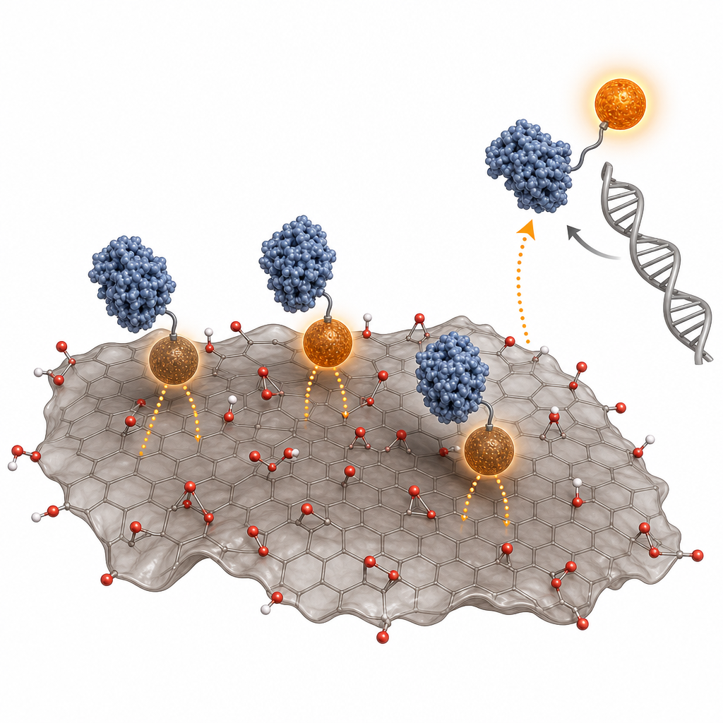
The ACS Material graphene oxide was central to the sensing mechanism. The Materials section states that the "GO dispersion (ACS Material, Pasadena, CA) was vortexed to ensure a homogeneous solution before diluting with deionized water." The stock dispersion was 5 mg/mL and was serially diluted from 1 mg/mL down to the microgram-per-milliliter working range. The product had a single-layer ratio greater than 80%, sheet sizes from 0.5 to 2.0 µm, and a thickness of 0-2 nm, characteristics confirmed by TEM and AFM. In the assay, 10 µL of 50 µg/mL GO dispersion was incubated with QD-labeled TALEs in TALE storage buffer for 30 minutes, after which target dsDNA was added. The large, biocompatible GO basal plane allowed the TALE proteins to adsorb through non-covalent interactions such as π-π stacking, electrostatic interaction, and hydrogen bonding, bringing the quantum dots within the ~30 nm FRET quenching range. The authors quantified GO quenching efficiency across 0-10 µg/mL and selected 5 µg/mL as the optimum.
Key quantitative results demonstrate the platform's sensitivity and specificity. At 5 µg/mL, GO produced approximately 28% quenching of QD-labeled TALEs, matching the ~30% optimum predicted for a large 120 kDa protein. TEM confirmed ~10 nm QDs adsorbed on GO, and AFM measured an adsorbed QD-TALE height of about 20 nm, consistent with FRET quenching within 30 nm. Two engineered TALEs were tested: tetM_1298 (recognizing 14 base pairs) and tetM_611 (16 base pairs), with EMSA-measured binding affinities (kD) of 1.2 and 4.6 nM, respectively. Using DNA oligonucleotides, the limit of detection was 10 pM for tetM_1298 and 1 pM for tetM_611, with fluorescence recovery scaling quantitatively from 1 pM to 100 nM. With S. aureus genomic DNA containing tetM, tetM_611 reached a 1 fM detection limit (equivalent to ~1.8 pg/µL), while tetM_1298 reached 10 fM. The longer 16 bp recognition site of tetM_611 conferred higher sensitivity, attributed to a more stable TALE-DNA complex. The assay also distinguished target from nontarget and irrelevant DNA sequences, confirming high specificity, and required only a 10-minute DNA incubation at room temperature.
This work enables rapid, equipment-light screening of antibiotic resistance genes without PCR or DNA labeling, with clear relevance to point-of-care diagnostics, clinical microbiology, and surveillance of resistance in environmental and agricultural samples. Because TALEs are fully programmable, the same GO-FRET architecture can be extended to multiplexed detection by labeling distinct TALEs with different-colored quantum dots to recognize multiple resistance genes simultaneously. The authors note that integrating the assay into a microfluidic module could further lower the detection limit and that future work will focus on real-world biological samples such as cell lysate. The biocompatibility and broad quenching range of graphene oxide make it a versatile transducer for protein- and nucleic-acid-based optical sensors.
For researchers building similar FRET-based biosensors, the graphene oxide dispersion used here is available from ACS Material in the company's Graphene Series. The paper shows that a well-dispersed, predominantly single-layer GO with controlled sheet size and thickness performs reliably as a fluorescence quencher and protein adsorption surface, supporting reproducible detection across pM-to-fM target concentrations. This makes high-quality GO dispersions a practical building block for diagnostic, environmental, and bioanalytical platforms that rely on tunable fluorescence quenching.How ACS Material products were used
- Graphene Oxide (GO) Dispersion (5 mg/mL, single-layer ratio >80%, sheet size 0.5-2.0 µm, thickness 0-2 nm) (Graphene Series) — “GO dispersion (ACS Material, Pasadena, CA) was vortexed to ensure a homogeneous solution before diluting with deionized water. ... The single layer ratio is >80% with the size of GO sheets ranging from 0.5 to 2.0 μm and a thickness of 0-2 nm.”
Product Performance in this StudyThe ACS Material graphene oxide dispersion served as the FRET fluorescence quencher and adsorption platform for QD-labeled TALEs, enabling a turn-on biosensor with a detection limit as low as 1 fM of genomic DNA. It performed as an effective quencher, giving ~28% quenching at 5 µg/mL.
Related product categories
Frequently asked questionsHow does graphene oxide improve fluorescence biosensor sensitivity?
Graphene oxide acts as a broad-band fluorescence quencher through fluorescence resonance energy transfer (FRET). When quantum-dot-labeled TALE proteins adsorb onto the GO surface, the quantum dot emission is quenched within roughly 30 nm. Upon binding target DNA, the probe dissociates and restores fluorescence, producing a turn-on signal. In this study, 5 µg/mL GO gave about 28% quenching and enabled detection down to 1 fM of genomic DNA.
What is graphene oxide used for in antibiotic resistance gene detection?
In this work graphene oxide served as both a fluorescence-quenching transducer and a protein adsorption surface. QD-labeled TALE proteins engineered to recognize the tetM resistance gene adsorbed onto GO nanosheets, quenching the quantum dots. Target double-stranded DNA binding released the probes and restored the signal, allowing direct, amplification-free detection of antibiotic resistance genes at room temperature in about 10 minutes.
Why is single-layer graphene oxide important for FRET-based sensors?
A high single-layer ratio and controlled sheet size ensure a large, uniform two-dimensional surface for protein adsorption and consistent fluorescence quenching. The GO used here had a single-layer ratio above 80%, sheet sizes of 0.5-2.0 µm, and a thickness of 0-2 nm, which kept quantum dots within the FRET range and supported reproducible, sensitive detection across pM-to-fM target concentrations.