A blue light transilluminator and a UV transilluminator do the same job: they light a stained gel from below so that DNA bands glow, but they reach the stain through different parts of its spectrum. UV units excite many stains in the ultraviolet, while blue-light viewers use visible blue light, with the gel viewed through an amber filter, and suit stains that absorb strongly in the blue [1][2]. This guide covers excitation and emission, both light sources, stain matching, DNA and operator exposure, documenting and gel cutting. For where gel viewing sits in the wider bench workflow, see From Extraction to Gel Viewing: A Molecular Biology Bench Workflow with PCR, qPCR and Electrophoresis.
Quick answer:
- Same job, different light: both light the gel from below; the cited gel-viewing literature includes UV-B units around 300–312 nm, while blue-light viewers use visible blue light and an amber filter [1–3].
- Match the stain: a stain must absorb well at the source wavelength; some cyanine stains absorb strongly near 495 nm, and one also near 300 nm [1][4].
- DNA in the gel: UV-B light damages stained DNA, which can spoil cloning or PCR from excised bands; blue-light viewing has been reported to cause much less damage [2][3].
- Not hazard-free: bright blue light has its own retinal blue-light hazard, so view through the amber filter or suitable eyewear and follow your institution's rules [5].
1. Excitation and Emission Basics
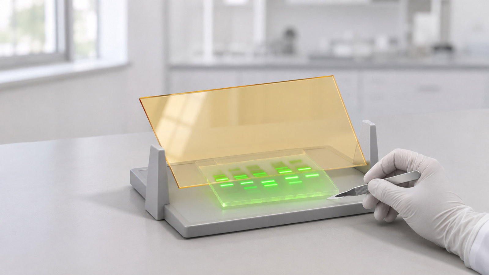
A fluorescent stain absorbs light in one or more bands of wavelengths and gives part of that energy back as light at longer wavelengths. A filter that blocks the excitation light but passes the emission lets the bands stand out against a dark background [2][6]. Binding to DNA changes the spectra: for ethidium bromide and propidium iodide, fluorescence rises about tenfold on binding and the excitation maxima move about 30–40 nm towards the red [7]. Shorter wavelengths also carry more energy per photon (§5).
Calculated illustration, not a specification. Inputs are hypothetical, not a recommended setting and not an ALab specification: photons at 300, 312 and 470 nm. From E = hc/λ, each photon carries about 4.13, 3.97 and 2.64 eV, so a 312 nm photon carries 470 ÷ 312 = 1.51 times the energy of a 470 nm photon. Damage also depends on how strongly DNA absorbs the light and on the dose [8].
2. UV Transilluminators
Standard protocols stain agarose gels with fluorescent intercalating dyes, such as ethidium bromide or a dual-excitation cyanine gel stain, and examine the gel directly in UV light; depending on the stain, bands containing as little as 20 pg of double-stranded DNA can be detected this way [1][9]. The cited gel-viewing literature includes UV-B units at 300, 306 and 312 nm [1][3]. ICNIRP places UV-B at 280–315 nm and UV-A at 315–400 nm [10].
UV still fits many laboratories: established ethidium methods are written for it, and a dual-excitation cyanine stain was developed with a maximum near 300 nm for laboratories equipped with 306 or 312 nm UV units alone [1][9]. The trade-offs are exposure of the DNA (§5) and of the operator: ICNIRP sets ultraviolet exposure limits for the eye and skin. For artificial sources it prefers engineering controls to protective clothing, eyewear and procedural measures [10]. Use the unit's shield or cover and follow your institution's rules for face and eye protection.
3. DNA Gel Viewing with Blue Light
Blue-light viewers swap the UV lamps for visible blue light and put an amber filter between the gel and the eye. The filter blocks the blue excitation light and passes the longer-wavelength emission, so the fluorescence is directly visible [2]. One description of the approach, using low-intensity broad-band blue light and broad-band filters, reports much less DNA damage than with a UV device, a benefit for downstream cloning, and minimal dye photobleaching [2]. The limit is the stain: one that absorbs mainly in the UV will look faint (§4).
| Feature | UV transilluminator (literature) | TI470 |
|---|---|---|
| Light source | UV-B lamps [1][3] | Bottom built-in LED light module |
| Wavelength | About 300–312 nm; 300, 306 and 312 nm units described [1][3] | Max emission wavelength 470 nm |
| Emission bandwidth | — | — |
| Stains | UV-excitable stains, including ethidium bromide [9] | Stains excited in roughly 450–495 nm |
| Viewing filter | — | Amber filter cover; rotates to several angles |
| Filter cut-off | — | — |
| Observation area | — | 210 × 120 mm |
Table 1. UV column: general literature, not a named product. — = not specified.
The ALab™ TI470 Blue Light Transilluminator is a blue-LED transilluminator (gel viewer) for the observation of nucleic acid and protein gel staining. The light comes from a bottom built-in LED light module; the maximum emission wavelength is 470 nm, and no emission bandwidth is specified. It is intended for stains excited in roughly the 450–495 nm range. That range describes the stains, not the light output. It uses an amber filter cover that blocks the blue excitation light so the stain's emission can be seen. The observation area is 210 × 120 mm, with an LED lifetime of more than 50,000 h, an intensity knob, and automatic shutdown after 5 min; the intensity setting is memorised. It runs from a 12 V, 1 A power adapter on AC 100–230 V, 50/60 Hz, measures 265 × 195 × 30 mm (L × W × H) and weighs 1 kg.
To check the configuration for your gels, see the ALab™ TI470 Blue Light Transilluminator: View product / request a quote.
4. Matching Stains to Light: 470 nm Gel Viewing
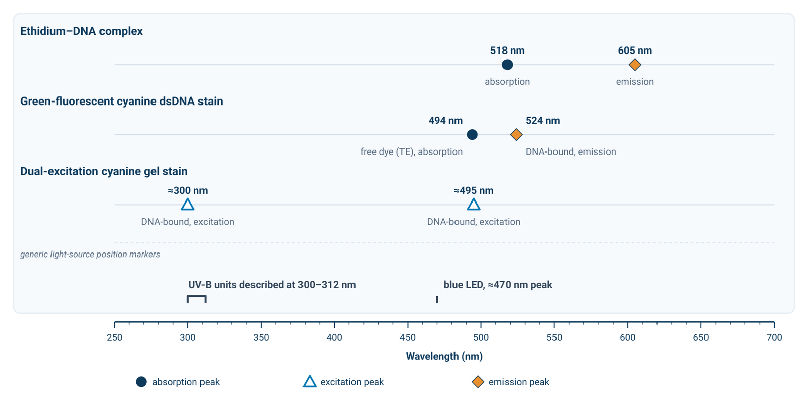
For 470 nm gel viewing, ask how strongly a stain absorbs near 470 nm and whether its emission is long enough to pass the amber filter. Figure 1 marks peak positions reported in the cited sources. For one green-fluorescent cyanine stain, the free dye has an absorption maximum at about 494 nm in TE buffer, while the dsDNA-bound complex emits at about 524 nm [4]. A dual-excitation cyanine gel stain has two excitation maxima when bound to DNA, at about 300 and about 495 nm [1]; the 495 nm peak indicates a visible-light excitation band, but whether a particular 470 nm viewer excites it adequately depends on the full spectra. The ethidium–DNA complex absorbs maximally at about 518 nm and emits at about 605 nm [7]. Peak positions alone do not predict signal strength; the full stain spectrum, source spectrum and filter transmission matter.
Stain choice can also affect sizing: in one comparison, two cyanine stains altered DNA mobility and therefore size estimates [11], so stain the ladder and samples the same way. Treat peak positions as a starting point and check the stain manufacturer's spectra before changing methods.
5. Sample and Operator Considerations
DNA in the gel. Light from a standard transilluminator fitted with 312 nm tubes damaged DNA in ethidium-stained gels. The recovered DNA was a poor substrate for in vitro transcription, transformation of E. coli and PCR, and adding cytidine or guanosine to the electrophoresis buffer protected it [3]. Earlier work reported similar damage from ethidium staining plus UV exposure [12]. Direct absorption of UV-B forms pyrimidine dimers, while UV-A photo-oxidation mostly targets guanine through singlet oxygen [8]. Blue-light viewing has been reported to cause much less damage [2].
Eyes and skin. Visible light is not hazard-free. ICNIRP describes a retinal blue-light hazard: photochemical injury, with a sensitivity that peaks at about 440 nm and absorption of 380–520 nm light by the retinal pigment epithelium. Injury can follow either an extremely bright light for a short time or a less bright light for longer [5][13]. IEC 62471 sets out exposure limits, reference measurement techniques and a classification scheme for lamps and lamp systems, including LEDs, from 200 to 3000 nm [14]. Part 7 of the series, IEC 62471-7:2023, specifies a photobiological safety assessment for light sources and luminaires that emit predominantly visible radiation (380–780 nm) [15]; this guide gives no assessment of any instrument. For blue-light viewing, keep the amber filter cover between the source and your eyes; for UV viewing, use the unit's shield or cover. In both cases, follow your institution's rules for suitable eye and face protection.
Stain handling. Handle every nucleic acid stain according to its manufacturer's data sheet; under illumination, the ethidium–DNA complex can also photobleach and cause single-strand breaks [7].
6. Viewing vs Documenting
Viewing means looking at the gel by eye to check a run or to find a band for cutting. Documenting means recording the gel with a camera or imager, which needs its own emission filter, exposure settings and a light source matched to the stain. Blue-light systems can serve both direct viewing and camera-based detection [2]. The TI470 is designed for viewing and gel cutting; it is a viewer, not a documentation system, so a recorded image needs a separate camera or imager.
7. Gel Cutting for Downstream Work
Stained DNA bands can be cut out and recovered from the gel [9]. Cutting takes longer than a quick look, so work quickly under UV, or use blue light with a stain that suits it (§4) to limit damage to the recovered DNA [2][3]. Use a clean scalpel or blade for each band, cut close to the band to keep excess agarose down, and switch the light off between cuts. On the TI470, the amber cover rotates to several angles for viewing and gel cutting. Gel percentage and fragment-size choices are covered in Agarose Gel Percentage and DNA Fragment Size: Choosing and Running a Horizontal Gel.
8. Frequently Asked Questions
Blue light vs UV transilluminator: which suits my gels?
It depends on the stain. UV suits stains excited mainly in the UV, including standard ethidium methods. Blue light suits stains that absorb strongly near 470–495 nm, and it is worth considering when bands will be cut for cloning or PCR because blue-light viewing has been reported to cause much less DNA damage than UV [2][3][4][9] (§4, §5).
Can I view ethidium bromide gels under blue light?
The ethidium–DNA complex has an absorption maximum at about 518 nm [7]. For the TI470, the maximum emission wavelength is 470 nm, and no full output spectrum or bandwidth is specified; those peak values alone do not establish whether a particular ethidium-stained gel will be visible. Signal depends on spectral overlap, the filter and stain concentration, so test your own gels [2][6][7]. Ethidium protocols are written for UV viewing [9] (§4).
Do I still need eye protection with blue light?
Follow your institution's rules. Bright blue light carries a retinal blue-light hazard, so view through the amber filter or suitable eyewear [5] (§5).
Does UV damage DNA I want to clone?
It can. DNA recovered from UV-B-illuminated gels was a poor substrate for transcription, transformation and PCR, and blue-light viewing has been reported to cause much less damage [2][3] (§5).
9. Related Guides
- From Extraction to Gel Viewing: A Molecular Biology Bench Workflow with PCR, qPCR and Electrophoresis
- Agarose Gel Percentage and DNA Fragment Size: Choosing and Running a Horizontal Gel