Cryo-electron microscopy has transformed structural biology,1 but specimen preparation remains a stubborn bottleneck. During conventional vitrification, protein particles can reach the air–water interface within milliseconds, where some denature or adopt preferred orientations.2,3 Graphene grids for cryo-EM provide an ultrathin, conductive support that can intercept particles before they spend prolonged time at that interface — but performance depends on transfer quality, surface cleanliness, and functionalization. This guide reviews the evidence: functionalized graphene supports, the EG-grid results, and a published batch workflow that produced 36 graphene-coated grids in 1.5 days using Trivial Transfer® Graphene as the transfer starting material.
Explore Trivial Transfer® Graphene
The Air–Water Interface Problem in Cryo-EM Sample Preparation
Advances in direct electron detectors and maximum-likelihood image processing carried single-particle cryo-EM into near-atomic territory — the shift celebrated as the resolution revolution.1 The part of the workflow that has advanced least sits earlier: the few microliters of sample pipetted onto the grid, and what happens to it in the second before vitrification.
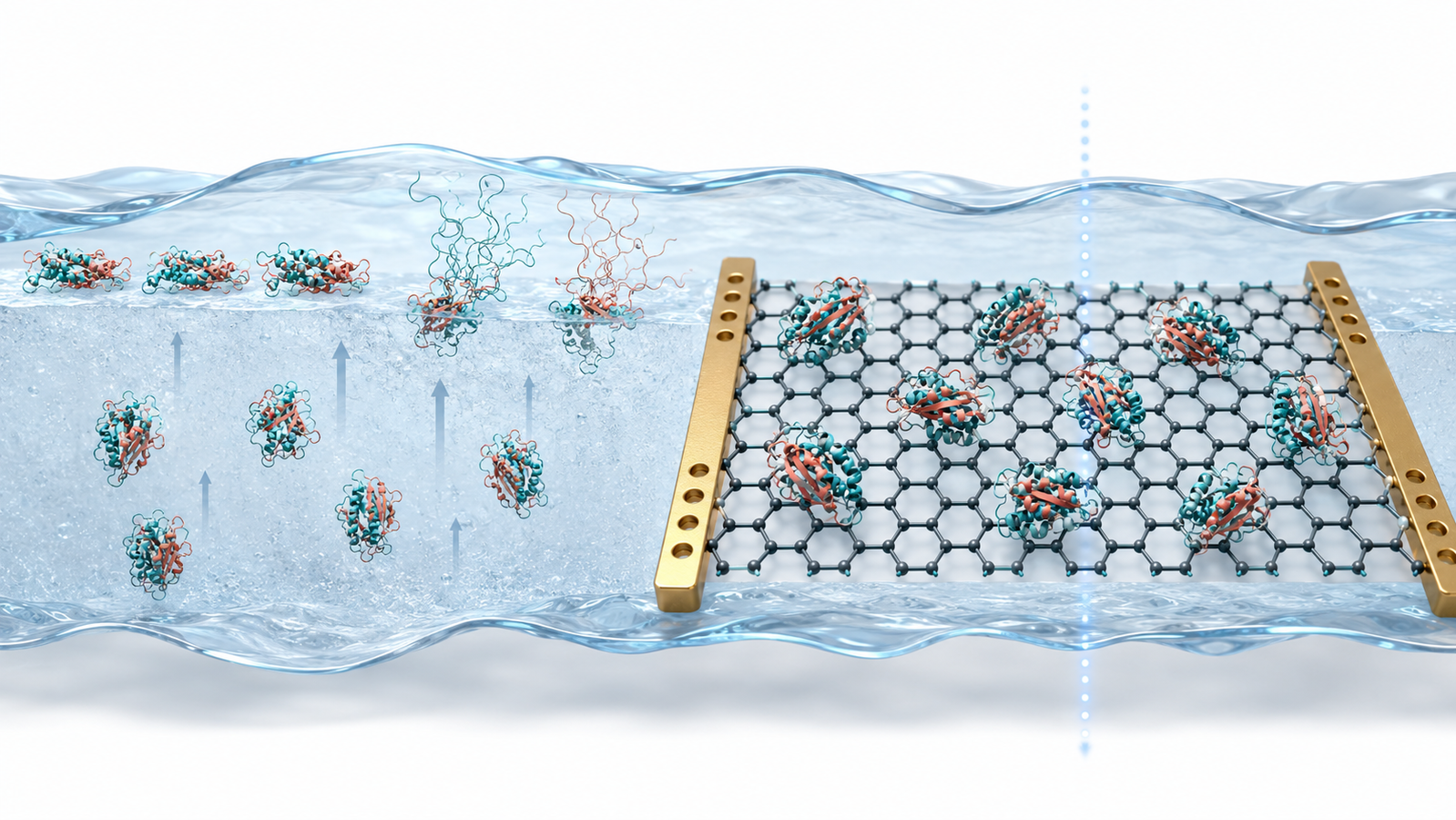
In conventional preparation, the specimen ends up as a vitrified film tens of nanometers thick, suspended across the holes of a fenestrated support. Inside that film, a protein molecule can diffuse to the air–water interface (AWI) in milliseconds — and tomographic surveys of real grids have shown that in the large majority of specimens examined, particles were adsorbed to that interface rather than floating free in the ice.2 The interface is not a neutral parking spot. Some particles can partially unfold there — denaturation at the AWI has been directly documented — while others survive intact but adopt a strongly preferred orientation, starving the reconstruction of the views it needs.3
The downstream symptoms are familiar in cryo-EM sample preparation: anisotropic maps that stretch along the missing views, long collections to harvest enough rare orientations, half-empty holes that waste microscope time, and sample-concentration demands the biochemistry cannot always meet. None of these are camera problems. They frequently begin at the interface.
Why Graphene Support Films Are Different
Giving the particle somewhere better to land is an old idea — amorphous carbon has played that role for decades — but conventional continuous carbon supports generally add more background scattering than a monolayer of graphene. Graphene changes the arithmetic for four reasons.
It is nearly invisible. A support one atom thick adds minimal additional background relative to conventional continuous carbon supports, so more of the surviving signal belongs to the protein. It is strong enough to be that thin. Monolayer graphene is among the strongest materials ever measured, which is what allows a single atomic layer to span micrometer-scale holes intact.4 It conducts, electrically and thermally, which can mitigate the charging and beam-induced motion that degrade high-resolution frames. And it intercepts the particle: adsorption to graphene can reduce prolonged exposure to the air–water interface.3,5
There is one catch, and much of the modern literature on functionalized graphene grids is a response to it: as-transferred graphene is hydrophobic. An aqueous protein sample may bead up instead of spreading, preventing the support from functioning reliably. How you make graphene wettable — and what chemistry you leave on its surface when you do — strongly influences which particles stick, at what density, and in which orientations.
Interactive: Interface vs Graphene Support
The schematic model below illustrates the argument of this article. Choose a grid type and watch where the particles tend to end up in the simulation: at the interface, beaded up on unwetted graphene, or adsorbed on a functionalized support — with a model orientation-coverage diagram updating live. It is an illustrative teaching model, not a predictor of experimental outcomes.
| Support type | Simulated tendency in the model | What the literature reports |
|---|---|---|
| Open hole (no support) | Higher simulated risk of interface adsorption and restricted orientation coverage | Particles adsorbed at the AWI in most surveyed specimens; denaturation documented in some cases2,3 |
| Plain graphene (as transferred) | Poor wetting may reduce usable particle density | Hydrophobic surface requires activation or functionalization before use6 |
| Functionalized graphene | Functionalization can increase adsorption and broaden orientation coverage; outcomes remain specimen-dependent | Reported gains in particle density and orientation distribution for the specimens studied7,8,9 |
Functionalizing Graphene Grids: From Wettability to Orientation Control
The first generation of graphene-grid work — call it Era 1 — treated hydrophobicity as an obstacle to be removed. Low-energy hydrogen plasma treatment showed that graphene could be rendered protein-friendly without destroying its lattice, putting practical graphene supports on the map.6 Multifunctional graphene supports extended the idea into a general specimen-support platform.5
Then the field noticed something more useful: surface chemistry is not just a wettability switch — it can act as a steering wheel. Amino- and PEG-amino-functionalized graphene oxide grids were reported to enrich particles and shield them from the interface.7 Grids functionalized with ligands carrying different charges could reshape the orientation distribution itself for the specimens tested, selecting which face of the molecule contacts the support.8 The question quietly changed from “how do we make graphene wet?” to “what do we want the surface to do?” — the reframing that set up both studies at the heart of this article. (Graphene-oxide chemistry starts from a different feedstock than CVD film — for that branch, see our single-layer graphene oxide dispersion.)
Epoxidized Graphene Grids: Reported Resolution and Precursor Storage
In 2023 — Era 2 in this story — a team spanning Osaka University's frontier-biosciences, pharmaceutical-sciences, and protein-research institutes together with JEOL published a different route to a functional graphene surface. Instead of plasma or aggressive wet chemistry, Fujita, Makino, Asahara and colleagues oxidized graphene with photoactivated ClO2• as a mild oxidant, then built further chemistry on top to produce an epoxidized graphene grid (EG-grid™) that actively immobilizes protein particles.9
Two reported results stand out. The first concerns storage — with an important distinction. What the study validated is the ClO2•-oxidized precursor grid: it retained its reactivity for at least three months under N2 at room temperature, after which the authors completed the five-minute ECH epoxidation step and obtained a 2.06 Å GroEL reconstruction. The study did not establish the same three-month shelf life for a fully epoxidized, ready-to-use EG-grid.9 Even so, a precursor that keeps for months and is finished by a five-minute step on the day of use is a meaningful practical advantage over activation methods whose effect decays within hours.6,9 The second reported result is efficiency, for the specimens studied:
| Specimen | Reported resolution | Micrographs used |
|---|---|---|
| GroEL | 1.99 Å | 504 |
| GAPDH | 2.16 Å | 241 |
| SARS-CoV-2 spike protein | 3.10 Å | 1,163 — from a 0.1 mg mL−1 sample |
Reconstructions reported for the EG-grid in Ref. 9; the authors attribute the improvements to increased particle density and a broadened orientation distribution under their conditions.
A sub-2 Å map from roughly five hundred micrographs — for these specimens, under the reported conditions — changes what an overnight session can accomplish. And the spike-protein result makes the concentration point concrete: 0.1 mg mL−1 sufficed in that study, which brings within reach specimens that are difficult to purify to the concentrations open-hole grids often demand.9
Batch Preparation: 36 Graphene-Coated Grids in 1.5 Days
A functionalized graphene grid that works in one lab is a result. A protocol a facility can run every week is a technology — and between the two sits the step nobody writes headlines about: making enough grids, reproducibly, without heroics. That is Era 3.
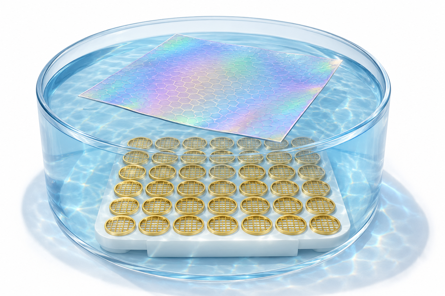
This gap is what Ahn and colleagues at the University of Michigan addressed in ACS Nano, with a reliable, easily implemented method that produced 36 graphene-coated grids at once within 1.5 days.10 The workflow is deliberately ordinary. A 25.4 × 25.4 mm PMMA-supported CVD graphene pad — the paper's methods record that the pad “(Trivial Transfer Graphene) was purchased from ACS Material” and stored refrigerated at 2–8 °C before use — is floated on deionized water above 36 gold Quantifoil grids arranged on a 3D-printed transfer tool, then lowered so the film settles across all of them in a single operation; the transfer procedure itself was based on the product's user instructions, with modifications.10
Quality control was built in rather than assumed: the grids were characterized by scanning electron microscopy (coverage and tears), Raman spectroscopy (layer number and defect density), and atomic force microscopy (surface topography).10 The biological validation was a head-to-head on the same specimen: the structure of Methylococcus capsulatus soluble methane monooxygenase hydroxylase was reported at 2.9 Å on a standard Quantifoil grid and 2.5 Å on the graphene-coated grid, with the authors noting that in their hands the graphene support needed less protein, made ice thickness easier to control, and helped keep the enzyme away from air–water-interface denaturation — differences sharp enough to reveal subtle geometry at the enzyme's non-heme diiron active site relative to the crystal structure.10
View Trivial Transfer® Graphene
Published Cryo-EM Applications of Graphene Supports
The surest sign a specimen technology is maturing is that it stops being the subject of the paper and moves into the methods section. Recent studies show graphene supports appearing in Methods sections across selected applications — and the three below all name the same commercial starting film. Structural studies whose specimen preparation used ACS Material Trivial Transfer Graphene span three of the categories that suffer most from interface pathologies:
- A human autophagy-initiating supercomplex — the ULK1C:PI3KC3-C1 assembly, structure and activation resolved in Nature Structural & Molecular Biology.11
- A voltage-gated potassium channel in two functional states — Kv1.2 captured conducting and non-conducting, in eLife.12
- A mitochondrial import complex straight from tissue — an ex vivo Drosophila TOM complex, in IUCrJ.13
Membrane channels, tissue-derived assemblies, multi-subunit supercomplexes — specimens where yield is scarce and orientations often misbehave. For a walkthrough of another published example on this site, see our review of a plant respiratory chain supercomplex study using a graphene support.
A Practical Graphene-Grid Workflow and Quality-Control Checklist
Start from a transferable film, not a pre-coated grid. Both anchor studies begin with PMMA-supported CVD graphene floated on water — a geometry that lets you pick your own grid (gold or copper, any mesh), batch at your own scale, and functionalize on your own schedule.9,10 Batch the transfer. The per-grid setup burden falls substantially when transfer is performed as a batch10 — the additional setup required for one-at-a-time transfer may contribute to the perception that graphene grids are difficult to prepare. Choose functionalization for your timeline. Plasma-style activation is fast but fades within hours; the epoxide route is more involved, but its oxidized precursor grids stored for months under nitrogen in the reported tests, with a five-minute epoxidation completed at time of use — the right choice depends on whether you prepare grids the day of, or the quarter before.6,9 Verify the film before spending protein. Raman for layer number and defects, SEM for coverage, AFM for topography — the published QC trio.10 And store the pad cold: the published protocol keeps it at 2–8 °C until use.10
When Graphene Grids May — and May Not — Be Necessary
An honest guide should say so: a graphene support is not the default answer for every specimen. When sample is plentiful and behaves well on a conventional holey grid — good particle density, adequate orientation spread, stable ice — the conventional grid is often the simpler choice. Graphene transfer adds real requirements of its own: film cleanliness and coverage control, polymer-residue removal, functionalization chemistry, and in-house QC capability. A graphene support earns its place when the specimen gives you a reason: sample scarcity, persistently low particle density, strong preferred orientation, suspected sensitivity to the air–water interface,2,3 or beam-induced-motion concerns — weighed against your lab's transfer and QC capacity.
Using Trivial Transfer® Graphene as the Transfer Starting Material
Trivial Transfer® Graphene is a PMMA-supported CVD graphene film that can serve as the transfer starting material for graphene-coated cryo-EM grids. It is not a finished, ready-to-vitrify grid: the cited workflows still require transfer onto the selected grid, PMMA removal and cleaning, surface activation or functionalization, and quality control.9,10 Its advantage is that researchers can begin with a transferable film and avoid growing graphene, spin-coating PMMA, and etching copper before transfer — the published batch workflow above started from exactly this film, following the product's transfer instructions.10 Researchers building specimen supports from the oxide branch instead can start from our single-layer graphene oxide water dispersion.
Explore Trivial Transfer® Graphene
FAQ
1. Why do cryo-EM samples need a support film at all?
Because in an open hole the protein can reach the air–water interface within milliseconds, and surveys of real grids show most particles ended up adsorbed there — where some unfold or lock into a preferred orientation.2,3 A support film gives the particle an alternative surface to land on first.
2. Is Trivial Transfer® Graphene a ready-to-use cryo-EM grid?
No. It is a PMMA-supported, transferable graphene film used as a starting material. Preparing a cryo-EM grid still requires transfer onto your chosen grid, PMMA removal, cleaning, surface treatment or functionalization, and quality control.9,10
3. What makes graphene different from amorphous carbon as a support?
Thickness and conductivity. A one-atom layer adds minimal additional background relative to conventional continuous carbon supports; its exceptional strength enables an atomically thin continuous support to span micrometer-scale holes,4 and its conductivity can mitigate charging and beam-induced motion.5
4. Why does graphene have to be functionalized first?
As-transferred graphene is hydrophobic — aqueous sample beads up on it. Functionalization (hydrogen plasma, graphene-oxide chemistries, or the epoxide route) makes the surface wettable, and the chemistry chosen also influences which particles adsorb and in what orientations.6,7,8,9
5. How many grids can one transfer pad make?
The published batch protocol reports coating 36 Quantifoil grids from a single 25.4 × 25.4 mm Trivial Transfer Graphene pad, in about a day and a half including quality control.10
6. When should I consider a graphene support — and when is a conventional grid simpler?
Consider graphene when sample is scarce, particle density is persistently low, orientations are strongly preferred, or the specimen appears sensitive to the air–water interface.2,3 When sample is plentiful and behaves well on a conventional holey grid, the conventional grid is often the simpler, faster choice — graphene adds transfer, cleanliness, and QC requirements of its own.
7. Does a graphene support guarantee higher resolution?
No. Resolution depends on specimen quality, graphene coverage and cleanliness, functionalization, ice thickness, vitrification, data acquisition, and processing. What the published head-to-head shows is that a well-prepared graphene support does not necessarily limit resolution: the same enzyme was reported at 2.5 Å on a graphene-coated grid versus 2.9 Å on a standard grid, with less protein consumed,10 and sub-2 Å reconstructions have been reported on functionalized graphene for other specimens.9
References
This article is provided by ACS Material LLC for educational purposes. Results, resolutions, and protocol details summarized here belong to the cited publications and their authors and were obtained under those studies' specific specimens and conditions; citation does not imply endorsement of ACS Material by those authors or their institutions, and published results do not guarantee equivalent outcomes with any product. The interactive model is a schematic teaching tool reflecting qualitative behavior described in the cited literature — its particle counts and probabilities are illustrative, not measured kinetics or predictions. Figures are AI-generated representative illustrations, not micrographs or artwork from the cited studies.